. 2000 Dec 1;19(23):6465-74.
doi: 10.1093/emboj/19.23.6465.

Immobilization of the early secretory pathway by a virus glycoprotein that binds to microtubules

Affiliations

Immobilization of the early secretory pathway by a virus glycoprotein that binds to microtubules

A Xu et al. EMBO J. .

Abstract

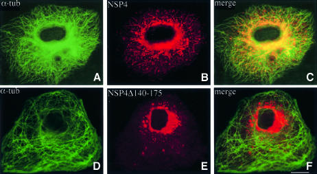
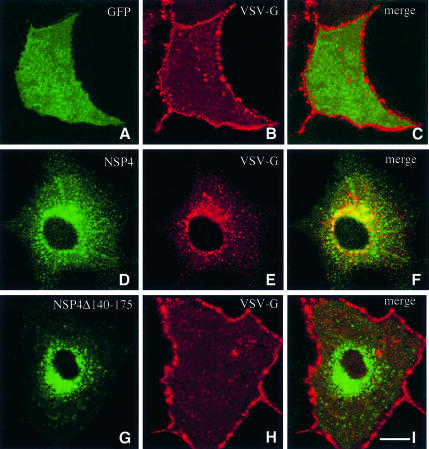
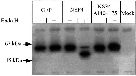
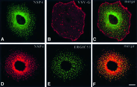
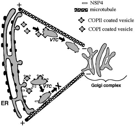
Membrane trafficking from the endoplasmic reticulum (ER) to the Golgi complex is mediated by pleiomorphic carrier vesicles that are driven along microtubule tracks by the action of motor proteins. Here we describe how NSP4, a rotavirus membrane glycoprotein, binds to microtubules and blocks ER-to-Golgi trafficking in vivo. NSP4 accumulates in a post-ER, microtubule-associated membrane compartment and prevents targeting of vesicular stomatitis virus glycoprotein (VSV-G) at a pre-Golgi step. NSP4 also redistributes beta-COP and ERGIC53, markers of a vesicular compartment that dynamically cycles between the ER and Golgi, to structures aligned along linear tracks radiating throughout the cytoplasm. This block in membrane trafficking is released when microtubules are depolymerized with nocodazole, indicating that vesicles containing NSP4 are tethered to the microtubule cytoskeleton. Disruption of microtubule-mediated membrane transport by a viral glycoprotein may represent a novel pathogenic mechanism and provides a new experimental tool for the dissection of early steps in exocytic transport.


Figures

None
Fig. 1. NSP4 interacts with a 50 kDa cellular protein. (A) Immuno precipitation of labelled proteins from MA104 cells. Cells were 35S-labelled for 6 h and chased for 1 h prior to infection (lanes 1 and 2) or mock infection (lane 3) with SA11 rotavirus. Cells were harvested at 7 h post-infection and lysates immunoprecipitated with anti-NSP4 mAb (lanes 1 and 3) or pre-immune serum (lane 2). (B) Immunoprecipitation of labelled cellular proteins from transfected Cos-7 cells. Cells were transfected with pcNSP4 (lanes 4 and 5) or pCDNA3.1 (lane 6) and 35S-labelled for 6 h commencing 40 h after transfection. Lysates were immunoprecipitated with anti-NSP4 mAb (lanes 4 and 6) or pre-immune serum (lane 5). Arrows indicate the positions of glycosylated (upper) and unglycosylated (lower) NSP4. (C) Pull-down assay using the C-terminal 90 amino acids of NSP4 (C90) fused to GST as bait. The procedure is described in Materials and methods. The specificity of the interaction was tested by addition of excess C90 (100 µg/ml) (lane 8). Proteins were resolved by 12% SDS–PAGE and visualized by autoradiography. *, the 50 kDa cellular protein identified in each experiment.
None
Fig. 2. Purification and separation of the NSP4-associated cellular protein. (A) Cellular proteins were purified using immobilized GST–C90 (lane 1) or GST as a control (lane 2) as described in Materials and methods. Bound proteins were eluted with 500 mM NaCl and analysed by SDS–PAGE (12%) and Coomassie staining. (B) The eluted proteins shown in lane 1 were analysed further by two-dimensional gel electrophoresis. Note that the NSP4-associated cellular protein was resolved into two separate polypeptides (arrowheads).
None
Fig. 3. The microtubule-binding domain of NSP4 is located within the C-terminal 54 amino acids. (A) Schematic representation of the structure of truncated variants of NSP4 used; H1–H3 denotes the positions of hydrophobic domains. (B) Cleared lysates from 35S-labelled MA104 cells were incubated with the indicated fusion protein immobilized on glutathione–agarose. Beads were recovered, washed, and bound proteins were detected by SDS–PAGE and autoradiography. C90* denotes a mutant form of C90 with a C-terminal methionine to isoleucine substitution previously shown to abolish binding of the rotavirus inner capsid particle (Taylor et al., 1993).
None
Fig. 4. Binding of 32P-labelled C90 to taxol-polymerized microtubules. Experiments were carried out as described in Materials and methods. Data points represent the mean and SEM from four independent experiments.
None
Fig. 5. Localization of NSP4 and NSP4Δ140–175 in vivo. Cos-7 cells were transiently transfected with a construct encoding either NSP4 or NSP4Δ140–175. After 48 h, the cells were fixed, permeabilized, and double-labelled with α-tubulin mAb (A and D) and a polyclonal antibody against NSP4 (B and E) prior to analysis by confocal laser scanning microscopy. Note the co-localization of α-tubulin with NSP4 (C), but not with the truncated mutant (F). The scale bar represents 10 µm.
None
Fig. 6. Expression of NSP4 blocks targeting of VSV-G to the plasma membrane. Cos-7 cells were transiently transfected with a construct encoding NSP4 (DF) or NSP4Δ140–175 (GI) and grown for 36 h. Cells were then re-transfected with in vitro synthesized mRNA encoding VSV-G and grown for a further 10 h. Cells were fixed, permeabilized, and double-labelled with a polyclonal antibody against VSV-G (B, E and H) and anti-NSP4 mAb (D and G). As a control, Cos-7 cells were transfected first with a construct encoding GFP (AC) prior to transfection of VSV-G mRNA. The scale bar represents 10 µm.
None
Fig. 7. Endo H sensitivity of VSV-G synthesized in the presence of NSP4 or NSP4Δ140–175. Cells were transfected as described in the legend to Figure 5. The cells were 35S-labelled for 2 h commencing 45 h post-DNA transfection and chased for a further 1 h prior to harvest. Lysates were immunoprecipitated with anti-VSV-G antiserum and half of the sample incubated with endo H. Proteins were visualized by SDS–PAGE and autoradiography.
None
Fig. 8. Distribution of ERGIC53, β-COP and Sec13p in cells expressing NSP4. Cos-7 cells were transiently transfected with pcNSP4 (B–D, F–H and J–L) or mock transfected (A, E and I) and grown for 48 h. Cells were fixed, permeabilized and double-labelled with a polyclonal antibody against NSP4 and a monoclonal antibody against either ERGIC53 (AD), β-COP (EH) or Sec13p (IL) prior to analysis by confocal laser scanning microscopy. The scale bar represents 10 µm.
None
Fig. 9. Distribution of NSP4, VSV-G and ERGIC53 in nocodazole-treated cells. Cells were transfected with either pcNSP4 and VSV-G mRNA as in Figure 5 (AC) or pcNSP4 alone (DF). At 48 h post-transfection, the cells were treated with 5 µM nocodazole for 3 h and then double-labelled for VSV-G and NSP4 (A–C) or ERGIC53 and NSP4 (D–F). The scale bar represents 10 µm.
None
Fig. 10. Schematic representation of NSP4-mediated arrest of ER-to-Golgi transport. Normal ER-to-Golgi translocation of VTCs is depicted along the upper microtubule. The presence of NSP4 causes a direct attachment of VTCs to the microtubule surface and prevents subsequent translocation.

References

    1. Andersson H., Kappeler,F. and Hauri,H.P. (1999) Protein targeting to endoplasmic reticulum by dilysine signals involves direct retention in addition to retrieval. J. Biol. Chem., 274, 15080–15084. - PubMed
    1. Ball J.M., Tian,P., Zheng,C.Q., Morris,A.P. and Estes,M.K. (1996) Age-dependent diarrhea induced by a rotaviral nonstructural glycoprotein. Science, 272, 101–104. - PubMed
    1. Bannykh S.I., Nishimura,N. and Balch,W.E. (1998) Getting into the Golgi. Trends Cell Biol., 8, 21–25. - PubMed
    1. Barlowe C. (1998) COPII and selective export from the endoplasmic reticulum. Biochim. Biophys. Acta, 1404, 67–76. - PubMed
    1. Bergmann C.C., Maass,D., Poruchynsky,M.S., Atkinson,P.H. and Bellamy,A.R. (1989) Topology of the non-structural rotavirus receptor glycoprotein NS28 in the rough endoplasmic reticulum. EMBO J., 8, 1695–1703. - PMC - PubMed

Publication types

MeSH terms