. 2000 Dec 1;19(23):6475-88.
doi: 10.1093/emboj/19.23.6475.

Nud1p links astral microtubule organization and the control of exit from mitosis

Affiliations

Nud1p links astral microtubule organization and the control of exit from mitosis

U Gruneberg et al. EMBO J. .

Erratum in

  • EMBO J 2001 Jan 15;20(1-2):305

Abstract

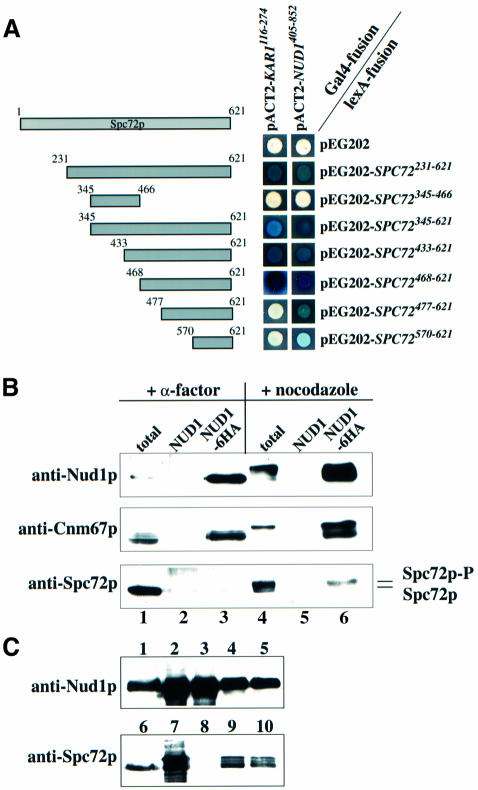
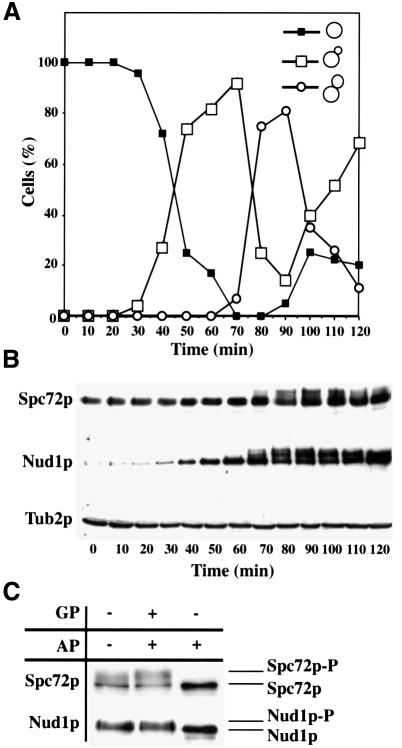
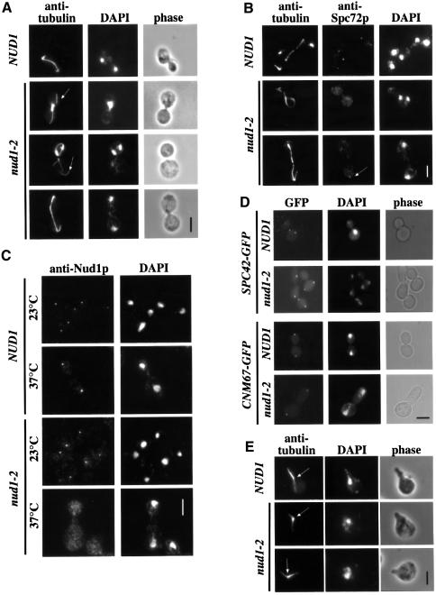
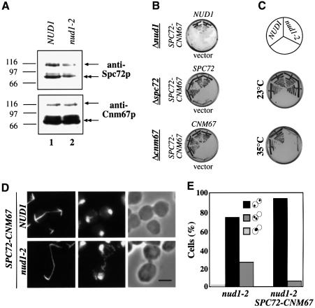
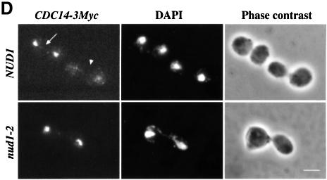
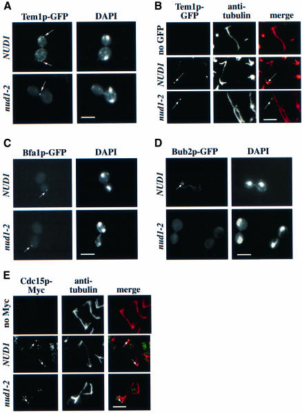
The budding yeast spindle pole body (SPB) not only organizes the astral and nuclear microtubules but is also associated with a number of cell-cycle regulators that control mitotic exit. Here, we describe that the core SPB component Nud1p is a key protein that functions in both processes. The astral microtubule organizing function of Nud1p is mediated by its interaction with the gamma-tubulin complex binding protein Spc72p. This function of Nud1p is distinct from its role in cell-cycle control: Nud1p binds the spindle checkpoint control proteins Bfa1p and Bub2p to the SPB, and is part of the mitotic exit network (MEN) in which it functions upstream of CDC15 but downstream of LTE1. In conditional lethal nud1-2 cells, the MEN component Tem1p, a GTPase, is mislocalized, whereas the kinase Cdc15p is still associated with the SPB. Thus, in nud1-2 cells the failure of Tem1p to interact with Cdc15p at the SPB probably prevents mitotic exit.


Figures

None
Fig. 1. Spc72p interacts with Nud1p. (A) Spc72p and Nud1p interact in the yeast two-hybrid system. Fragments of SPC72 subcloned into plasmid pEG202 were co-transformed into yeast strain SGY37 with pACT2 containing either KAR1 (codons 116–274) or NUD1 (codons 405–852). (B) Spc72p co-precipitates with Nud1p. NUD1 (lanes 2 and 5) and NUD1-6HA (lanes 1, 3, 4 and 6) cells were grown to early logarithmic phase and arrested with either α-factor or nocodazole. Nud1p-6HA was immunoprecipitated using anti-HA beads. Spc72p and Cnm67p in the immunoprecipitates were detected by immunoblotting. Lanes 1 and 4 show the total extract before immunoprecipitation, and lanes 2, 3, 5 and 6 the immunoprecipitates. Lanes 1–3 were loaded with four times more extract than lanes 4–6 to make up for the fact that NUD1 is expressed at much lower levels in pheromone-treated cells (Figure 2B). (C) Insect cells were transfected with either SPC72 baculovirus, NUD1-His6 baculovirus or both. Cells were harvested 72 h post-infection, lysed and incubated with TALON beads. Shown are yeast extracts (lanes 1 and 6), total lysates (lanes 2, 4, 7 and 9) and TALON-precipitates (lanes 3, 5, 8 and 10) of cells transfected with NUD1-His6 (lanes 2 and 3), SPC72 (lanes 7 and 8), and SPC72 and NUD1-His6 (lanes 4, 5, 9 and 10). Note that no protein was detected by the anti-Nud1p or anti-Spc72p antibodies in untransfected insect cells (not shown).
None
Fig. 2. Spc72p and Nud1p are phosphorylated in a cell-cycle-dependent manner. Samples of α-factor-synchronized cells were taken every 10 min and analysed for (A) budding index and (B) Spc72p, Nud1p and β-tubulin by immunoblotting. β-tubulin was used as loading control. (C) Spc72p-3HA or Nud1p-6HA was precipitated from cell extracts and treated without any addition, with alkaline phosphatase (AP) or alkaline phosphatase and β-glycerolphosphate (GP). Samples were analysed by immunoblotting.
None
Fig. 3. Overexpression of C-NUD1 interferes with AM organization and cell-cycle progression. (A) Cells of SPC72–GFP (UGY151, lanes 1 and 3) and NUD1–GFP (UGY153, lanes 2 and 4) with Gal1-HA-C-NUD1 were grown to early logarithmic phase in medium containing 3% raffinose. Galactose (4%) was then added to induce the Gal1 promoter. Samples were taken before (lanes 1 and 2) and 6 h after addition of galactose (lanes 3 and 4) and analysed by immunoblotting with anti-HA antibodies for the expression of HA-C-NUD1. Markers are bovine serum albumin (BSA) (66 kDa) and ovalbumin (45 kDa). (B) Yeast cells (YPH499) were transformed with vector p416-Gal1 (sector 1), p416-Gal1-N-NUD1 (sector 2) and p416-Gal1-C-NUD1 (sector 3) and grown on either glucose or galactose plates for 2 days at 30°C. (C) Diploid wild-type cells (FY1679, sector 1) or diploid cells containing one chromosomal copy of Gal1-NUD1 (UGY194, sector 2) were grown as in (B). (D) Cells of (A) were analysed for SPB-localized GFP fluorescence or (E) processed for indirect anti-tubulin immunofluorescence. DNA was stained with DAPI. In order to facilitate comparisons, control cells (–Gal) in anaphase were selected. The arrows indicate detached microtubules. (D and E) Bars, 5 µm. (F) The number of GFP-labelled SPB signals was scored before and 6 h after the addition of galactose to cells of SPC72–GFP, NUD1–GFP and CNM67–GFP with Gal1-C-NUD1 (n = 200).
None
Fig. 4. Analysis of the phenotype of nud1-2 cells. (A–C) NUD1 and nud1-2 cells were shifted to 37°C for 3 h and then processed for (A) anti-tubulin, (B) anti-tubulin and anti-Spc72p, or (C) anti-Nud1p immunofluorescence. (A) Detached AMs are indicated by arrows. (B) Residual Spc72p-SPB staining can be observed in some nud1-2 cells (arrow). (D) Spc42p and Cnm67p localization are not affected in nud1-2 cells. SPC42–GFP NUD1 (UGY196), SPC42–GFP nud1-2 (UGY199), CNM67–GFP NUD1 (UGY198) and CNM67–GFP nud1-2 cells (UGY200) were shifted to 37°C for 3 h and inspected by phase contrast and fluorescence microscopy. (E) Pheromone-arrested nud1-2 cells have normal AM arrays at 37°C. NUD1 and nud1-2 cells were treated with α-factor for 2.5 h at 23°C and then shifted to 37°C for 30 min in the continued presence of α-factor. Microtubules were visualized using anti-tubulin antibodies. The arrows indicate the positions of the SPBs. Note also that nud1-2 cells shifted for only 30 min to 37°C showed a profound AM organization defect (not shown). (A–E) DNA was stained with DAPI. Bars, 5 µm.
None
Fig. 5. nud1-2 cells have a defective outer plaque. (A) NUD1 and (B) nud1-2 cells were incubated for 3 h at 37°C. Samples were prepared for thin section electron microscopy. (A) In NUD1 cells the cytoplasmic outer plaque (OP), the central plaque (CP) and AMs emanating from the outer plaque are clearly detectable. (B) In nud1-2 cells the outer plaque was misformed and reduced. AMs were associated with the half-bridge of some nud1-2 cells. AM, astral microtubules; CP, central plaque; HB, half-bridge; NE, nuclear envelope; NM, nuclear microtubules; OP, outer plaque. Bar, 120 nm.
None
Fig. 6. An SPC72–CNM67 gene fusion alleviates the nuclear migration defect of nud1-2 cells. (A) Cell extracts of SPC72–CNM67 NUD1 (UGY217; lane 1) or SPC72–CNM67 nud1-2 (UGY215; lane 2) cells were prepared and analysed by immunoblotting using anti-Spc72p or anti-Cnm67p antibodies. The arrows indicate the Spc72–Cnm67p fusion protein (100 kDa), the double arrows the endogenous Spc72p or Cnm67p. Markers: β-galactosidase (116 kDa), phosphorylase b (97 kDa) and BSA (66 kDa). (BSPC72–CNM67 complements for SPC72 or CNM67 but not for NUD1. Cells of Δnud1 pRS316-NUD1 (UGY111), Δspc72 pRS316-SPC72 (ESM448) or Δcnm67 pRS316-CNM67 (ESM431) were transformed with pRS315-NUD1, pRS315-SPC72 or pRS315-CNM67, as indicated, pRS315-SPC72–CNM67 or pRS315 (vector). Cells were grown at 23°C on 5-fluoroorotic (5-FOA) plates, which select against the URA3-based pRS316 plasmids. Growth indicates complementation. Transformants were tested in duplicate. (CSPC72–CNM67 does not rescue nud1-2 cells. Cells of SPC72–CNM67 NUD1 or SPC72–CNM67 nud1-2 were grown at 23 or 35°C for 3 days. (DSPC72–CNM67 NUD1 and SPC72–CNM67 nud1-2 cells were shifted to 37°C for 3 h and processed for indirect tubulin immunofluorescence. DNA was stained with DAPI. Bar, 5 µm. (E) Quantitative analysis of the nuclear migration defect and cell-cycle arrest of nud1-2 and SPC72–CNM67 nud1-2 cells of (D). Only large-budded cells (90% of the population) were counted. The dots within the symbolized cells indicate the position of the DAPI-staining regions.
None
Fig. 7. nud1-2 cells exhibit a phenotype similar to MEN mutants. α-factor-synchronized NUD1 and nud1-2 cells with CLB2-6HA or PDS1-6HA were released into 37°C pre-warmed medium. Samples were taken every 30 min and (A) analysed for DNA content by FACS analysis, (B) budding index or (C) Pds1p-6HA and Clb2p-6HA by immunoblotting. Tub2p was detected as loading control. (DNUD1 and nud1-2 cells carrying CDC14-3Myc were shifted to 37°C for 2 h and processed for indirect immunofluorescence using anti-Myc antibodies. DNA was stained with DAPI. The upper panel shows two large-budded wild-type cells, one of which has already released Cdc14p into the nucleus and cytoplasm (arrowhead) and the other one in which Cdc14p is still held in the nucleolus (arrow). Out of 200 large-budded nud1-2 CDC14-3Myc cells, only three were found in which Cdc14p had been released from the nucleolus. Cdc14p-3Myc was still retained in the nucleolus when nud1-2 cells were incubated for 3 or 4 h at 37°C. Bar, 5 µm.
None
Fig. 8. Localization of MEN components in nud1-2 cells. (A) Tem1p–GFP is mislocalized in nud1-2 mutants. TEM1–GFP NUD1 (UGY218) or TEM1–GFP nud1-2 cells (UGY201) were synchronized with α-factor and then shifted to 37°C. The cells were fixed after 105 min when most of the TEM1–GFP cells reached anaphase. The Tem1p–GFP signal was inspected by fluorescence microscopy. (B) Cells of (A) were analysed by anti-tubulin staining and confocal microscopy. In NUD1 cells, Tem1p–GFP co-localized with the SPB whereas in nud1-2 cells the Tem1p–GFP signal (green) was distinct from the tubulin staining (red). Note that the overlap of the green and red signals results in a yellow dot. (C) The Bfa1p–GFP signal is mislocalized in nud1-2 cells. Cells of BFA1–GFP NUD1 and BFA1–GFP nud1-2 were shifted to 37°C for 3 h and then analysed by fluorescence microscopy. (D) The Bub2p–GFP signal is lost in nud1-2 cells. Cells of BUB2–GFP NUD1 and BUB2–GFP nud1-2 were treated as in (C). (Enud1-2 cells in late anaphase exhibit only one SPB-associated Cdc15p-9Myc signal. NUD1 and nud1-2 cells carrying CDC15–9Myc were grown to early logarithmic phase. Cells were then shifted to 37°C for 3 h and processed for tubulin immunofluorescence. (A–E) The arrows indicate the GFP and 9Myc signals. When indicated, DNA was stained with DAPI. Bars, 5 µm.
None
Fig. 9. Bub2p interacts with Nud1p. (A) Two-hybrid interaction of Bub2p and Nud1p. (B) Wild-type (lanes 1 and 5), BUB2-3HA (lanes 2 and 6), TEM1-3HA (lanes 3 and 7) and BFA1-3HA (lanes 4 and 8) cells were arrested with nocodazole. Cell extracts were incubated with anti-HA antibodies bound to protein G beads. The total extracts (lanes 1–4) and immunoprecipitates (lanes 5–8) were tested for Nud1p (anti-Nud1p) and HA-tagged proteins (anti-HA) by immunoblotting.

Similar articles

Cited by

References

    1. Adams I.R. and Kilmartin,J.V. (1999) Localization of core spindle pole body (SPB) components during SPB duplication in Saccharomyces cerevisiae. J. Cell Biol., 145, 809–823. - PMC - PubMed
    1. Balasubramanian M.K., McCollum,D. and Surana,U. (2000) Tying the knot: linking cytokinesis to the nuclear cycle. J. Cell Sci., 113, 1503–1513. - PubMed
    1. Bardin A.J., Visintin,R. and Amon,A. (2000) A mechanism for coupling exit from mitosis to partitioning of the nucleus. Cell, 102, 21–31. - PubMed
    1. Brachat A., Kilmartin,J.V., Wach,A. and Philippsen,P. (1998) Saccharomyces cerevisiae cells with defective spindle pole body outer plaques accomplish nuclear migration via half-bridge-organized microtubules. Mol. Biol. Cell, 9, 977–991. - PMC - PubMed
    1. Byers B. and Goetsch,L. (1975) Behavior of spindles and spindle plaques in the cell cycle and conjugation of Saccharomyces cerevisiae. J. Bacteriol., 124, 511–523. - PMC - PubMed

Publication types

MeSH terms

Substances