. 2000 Dec 1;19(23):6558-68.
doi: 10.1093/emboj/19.23.6558.

Dynamic associations of heterochromatin protein 1 with the nuclear envelope

Affiliations

Dynamic associations of heterochromatin protein 1 with the nuclear envelope

N Kourmouli et al. EMBO J. .

Abstract

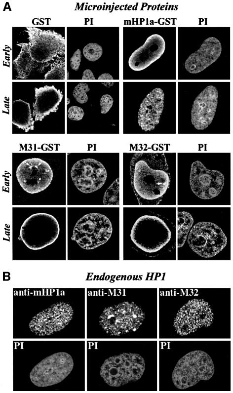
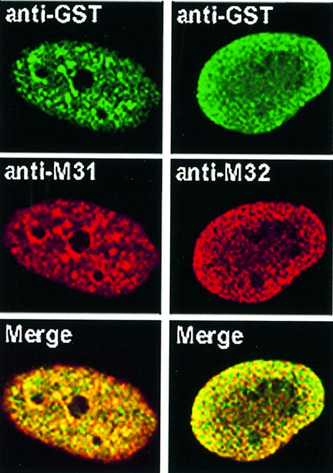
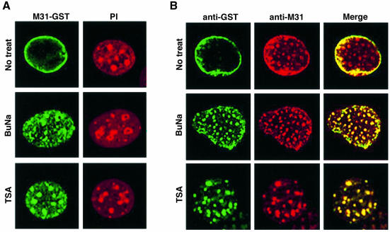
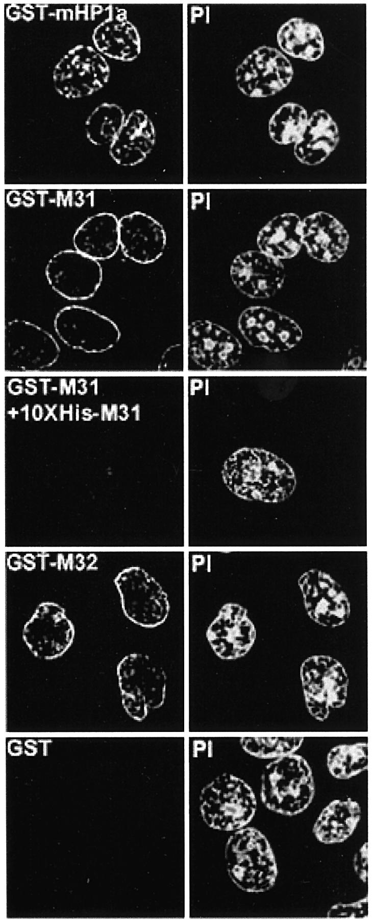
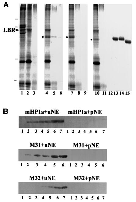
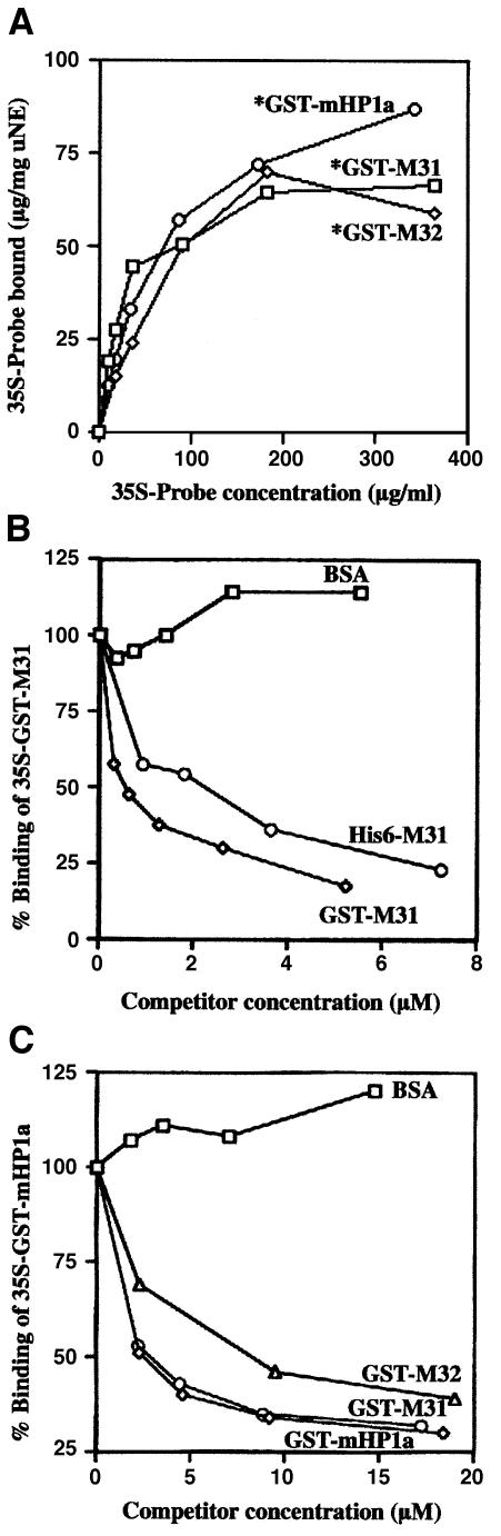
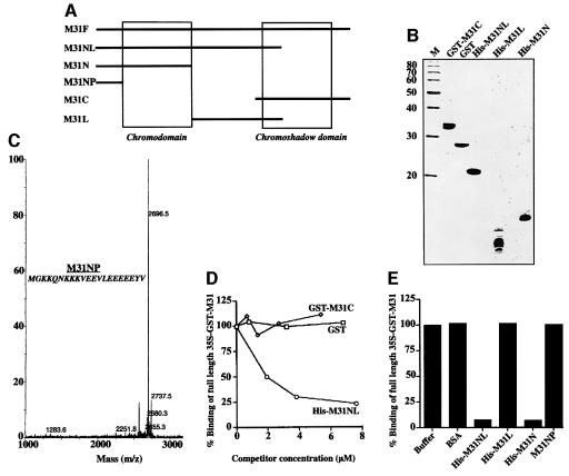
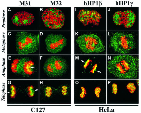
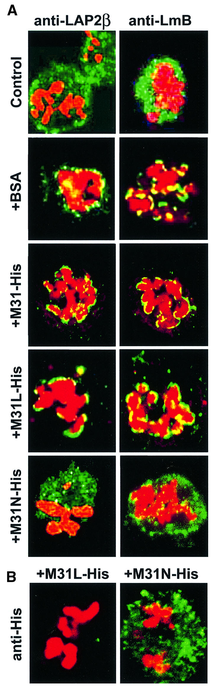
To study the dynamics of mammalian HP1 proteins we have microinjected recombinant forms of mHP1alpha, M31 and M32 into the cytoplasm of living cells. As could be expected from previous studies, the three fusion proteins were efficiently transported into the nucleus and targeted specific chromatin areas. However, before incorporation into these areas the exogenous proteins accumulated in a peripheral zone and associated closely with the nuclear envelope. This transient association did not occur when the cells were treated with deacetylase inhibitors, indicating an acetylation-inhibited interaction. In line with these observations, recombinant HP1 proteins exhibited saturable binding to purified nuclear envelopes and stained the nuclei of detergent-permeabilized cells in a rim-like fashion. Competition experiments with various M31 mutants allowed mapping of the nuclear envelope-binding site within an N-terminal region that includes the chromodomain. A His(6)-tagged peptide representing this region inhibited recruitment of LAP2beta and B-type lamins around the surfaces of condensed chromosomes, suggesting involvement of HP1 proteins in nuclear envelope reassembly.


Figures

None
Fig. 1. Distribution of exogenous HP1 proteins at various points after injection. (A) Characteristic distribution patterns of GST, GST–M31, GST–M32 and GST–mHP1α 5–20 (Early) and 40–120 min (Late) after injection into the cytoplasm of interphase HeLa cells. The recombinant proteins were detected with affinity-purified anti-GST antibodies. PI, the corresponding propidium iodide profiles. Arrows show foci of perinucleolar heterochromatin occasionally targeted by recombinant HP1 proteins. (B) Steady-state distribution of endogenous HP1 proteins in interphase HeLa cells. The specimens were stained with isotype-specific antibodies and propidium iodide (PI) as indicated.
None
Fig. 2. Structural features of recombinant M31. Circular dichroism spectra of purified His6-M31 (C), GST (D) and M31–GST (E).Curve F is the sum of the His6-M31 and GST spectra. The profile of fully denatured M31–GST is depicted in A. Plot B corresponds to the spectrum of the M31 peptide His6-M31N (for more information on this see Figure 8). The inset shows the electrophoretic mobility of the analyzed proteins in SDS–PAGE gels. Lane 1, molecular weight markers with values of 68, 41, 31 and 21 kDa; lane 2, pure GST; lane 3, M31–GST; lane 4, His6-M31.
None
Fig. 3. Co-localization of exogenous and endogenous HP1 proteins. Distribution of exogenous and endogenous M31 and M32 24 h after injection into the cytoplasm of HeLa cells. Staining with anti-GST antibodies appears in green, while staining with anti-M31 and anti-M32 antibodies (which detect both the endogenous and the exogenous products) appears in red. The merge shows the combination of the two patterns.
None
Fig. 4. Fate of microinjected M31–GST in cells treated with deacetylase inhibitors. (A) Patterns of M31–GST 5 h after injection into normal (No treat.), sodium butyrate-treated (BuNa) and TSA-treated (TSA) C127 cells. The recombinant protein was detected with anti-GST antibodies. PI, the corresponding propidium iodide profiles. (B) The same experiment as in (A) after double immunostaining with anti-GST and anti-M31 antibodies.
None
Fig. 5. In situ decoration of permeabilized cells by recombinant HP1 proteins. Triton X-100-treated cells (HeLa) after in vitro incubation with 40 µg/ml GST fusion proteins or a mixture of GST–M31 and His6-M31 in the molar ratio 1:10. Recombinant proteins were detected with affinity-purified anti-GST antibodies. PI, the corresponding propidium iodide profiles.
None
Fig. 6. Specific binding of HP1 proteins to isolated nuclear envelopes. (A) Binding of recombinant HP1 to isolated nuclear envelopes, as detected by SDS–PAGE and Coomassie Blue staining. Lane 1, whole nuclear envelopes (before urea extraction); lane 2, urea-stripped nuclear envelopes; lane 3, protease-digested nuclear envelopes; lane 4, urea-stripped nuclear envelopes plus GST–mHP1α; lane 5, protease-digested nuclear envelopes plus GST–mHP1α; lane 6, GST–mHP1α and buffer; lane 7, urea-stripped nuclear envelopes plus GST–M31; lane 8, protease-digested nuclear envelopes plus GST–M31; lane 9, GST–M31 and buffer; lane 10, urea-stripped nuclear envelopes plus GST–M32; lane 11, protease-digested nuclear envelopes plus GST–M32; lane 12, GST–M32 and buffer; lanes 13–15, profiles of the input GST–mHP1α, GST–M31 and GST–M32, respectively. Dashes correspond to molecular weight markers of 96, 68, 31 and 21 kDa. An arrowhead indicates the position of LBR. Dots indicate HP1 proteins that have bound to the membranes. (B) Binding of recombinant HP1 proteins to the nuclear envelopes, as detected by western blotting. The input was as follows: lanes 1, 0 µg; lanes 2, 0.5 µg; lanes 3, 1 µg; lanes 4, 2 µg; lanes 5, 4 µg; lanes 6, 8 µg; lanes 7, 16 µg. uNE and pNE correspond to urea-extracted and proteolyzed nuclear envelopes, respectively.
None
Fig. 7. Quantification of HP1 binding and competition experiments. (A) Concentration dependence of GST–mHP1a, GST–M31 and GST–M32 binding to urea-extracted nuclear envelopes. Metabolically labeled probes (signified by asterisks) were prepared as explained in Materials and methods. Background binding to proteolyzed membranes has been subtracted. (B and C) Competition experiments using radiolabeled probes (∼1 µM) and excess unlabeled HP1 proteins as indicated. All experiments shown here were done in triplicate. Variation between data points did not exceed 10%.
None
Fig. 8. The N-terminal region of M31 contains a nuclear envelope-binding site. (A) Schematic representation of the M31 mutants used in these experiments. The positions of the peptides relative to CD and CSD are indicated. (B) Purified proteins used in competition assays after SDS–PAGE and Coomassie Blue staining. M, molecular weight markers with the indicated molecular masses. (C) Sequence and mass spectra profile of the synthetic peptide M31NP, which was too small for analysis in regular SDS gels. (D and E) Competition of [35S]GST–M31 binding by various M31 mutants, GST and BSA (controls). All assays presented here were done in duplicate with ∼1 µM radiolabeled probe. Variation of data points was <10%.
None
Fig. 9. Localization of the three HP1 variants in mitotic cells. Mitotic C127 and HeLa cells doubly stained with anti-M31/M32 antibodies (green) and propidium iodide (red). Arrows show the localization of HP1 proteins to the polar sides of segregated chromosomes. Prophase, metaphase, anaphase and telophase cells are shown, as indicated.
None
Fig. 10. Effects of M31 peptides on nuclear envelope reassembly. Images labeled Control show permeabilized mitotic cells prior to incubation at 33°C. All other profiles correspond to samples that were incubated for 2 h at 33°C. (A) Assembly of nuclear envelope proteins assessed by staining the particles with anti-LAP2β or anti-lamin B antibodies (green) and propidium iodide (red). +BSA, bovine serum albumin; +M31-His, His6-tagged full-length M31; +M31L-His, a His6-tagged peptide containing residues 70–114 of M31; +M31N-His, a His6-tagged peptide containing residues 1–69 of M31. (B) Localization of the M31 peptides in ‘digitonin ghosts’ was determined after staining with anti-His6 antibodies.

Similar articles

Cited by

References

    1. Aagaard L. et al. (1999) Functional mammalian homologues of the Drosophila PEV-modifier Su(var)3-9 encode centrosome-associated proteins which complex with the heterochromatin component M31. EMBO J., 18, 1923–1938. - PMC - PubMed
    1. Aasland R. and Stewart,A.F. (1995) The chromo shadow domain, a second chromo domain in heterochromatin-binding protein 1, HP1. Nucleic Acids Res., 23, 3168–3174. - PMC - PubMed
    1. Ainsztein A.M., Kandels-Lewis,S.E., Mackay,A.M. and Earnshaw,W.C. (1998) INCENP centromere and spindle targeting: identification of essential conserved motifs and involvement of heterochromatin protein HP1. J. Cell Biol., 143, 1763–1774. - PMC - PubMed
    1. Ball L.J. et al. (1997) Structure of the chromatin binding (chromo) domain from mouse modifier protein 1. EMBO J., 16, 2473–2481. - PMC - PubMed
    1. Bodoor K., Shaikh,S., Salina,D., Raharjo,W.H., Bastos,R., Lohka,M. and Burke,B. (1999) Sequential recruitment of NPC proteins to the nuclear periphery at the end of mitosis. J. Cell Sci., 112, 2253–2264. - PubMed

Publication types

MeSH terms