. 2000 Dec 1;19(23):6569-81.
doi: 10.1093/emboj/19.23.6569.

Functional analysis of the human CDC5L complex and identification of its components by mass spectrometry

Affiliations

Functional analysis of the human CDC5L complex and identification of its components by mass spectrometry

P Ajuh et al. EMBO J. .

Abstract

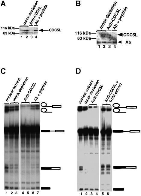
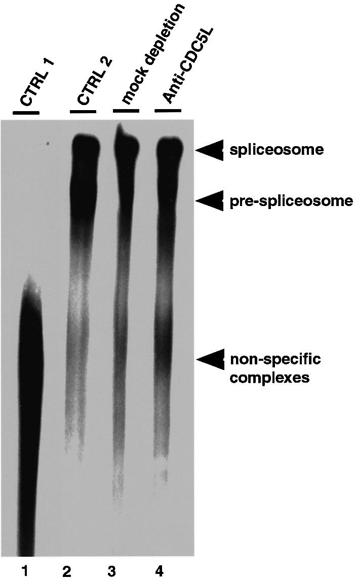
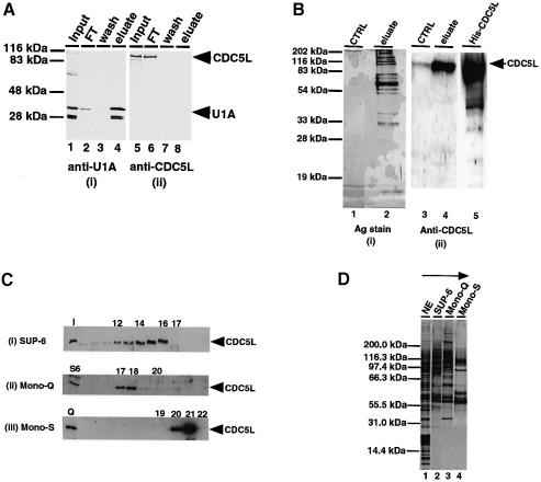
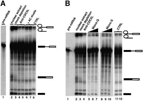
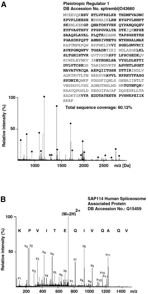
Recently, we identified proteins that co-purify with the human spliceosome using mass spectrometry. One of the identified proteins, CDC5L, corresponds to the human homologue of the Schizosaccharomyces pombe CDC5(+) gene product. Here we show that CDC5L is part of a larger multiprotein complex in HeLa nuclear extract that incorporates into the spliceosome in an ATP-dependent step. We also show that this complex is required for the second catalytic step of pre-mRNA splicing. Immunodepletion of the CDC5L complex from HeLa nuclear extract inhibits the formation of pre-mRNA splicing products in vitro but does not prevent spliceosome assembly. The first catalytic step of pre-mRNA splicing is less affected by immunodepleting the complex. The purified CDC5L complex in HeLa nuclear extract restores pre-mRNA splicing activity when added to extracts that have been immunodepleted using anti-CDC5L antibodies. Using mass spectrometry and database searches, the major protein components of the CDC5L complex have been identified. This work reports a first purification and characterization of a functional, human non-snRNA spliceosome subunit containing CDC5L and at least five additional protein factors.


Figures

None
Fig. 1. Specific immunoprecipitation of the spliceosome by anti-CDC5L. (A) pre-mRNA splicing reactions/spliceosomes were immunoprecipitated using 5–10 µg of anti-CDC5L antibody in the presence and absence of the antibody specific peptide (10-fold molar excess) and the immunoprecipitated RNA revealed by autoradiography. Lane 1 contained the pre-mRNA transcript used in the splicing reactions. Lanes 2 and 3 show the input splicing reaction. Lane 4 contains a mock immunoprecipitation using pre-immune IgG. Lanes 5 and 6 are duplicate immunoprecipitation reactions performed with anti-CDC5L antibodies in the presence of the antibody specific peptide. Lanes 7 and 8 are duplicate spliceosome immunoprecipitations using anti-CDC5L antibody. The symbols on the right of the figure represent the pre-mRNA band and the different splicing intermediates and products. All the other bands in the figure have been produced by RNA partial degradation. (B) Splicing reactions were prepared as above, but in the presence or absence of ATP and immunoprecipitation reactions performed using anti-CDC5L antibody. The amount of radioactivity immunoprecipitated was measured as relative fluorescence units (RFU) using a PhosphorImager (Molecular Dynamics) and the results presented on a bar chart. The bars marked 1, 3 and 4 represent the amount of radioactivity immunoprecipitated when the splicing reaction is carried out in the presence of ATP. However, in the case of bar 3, the immunoprecipitation was performed using anti-CDC5L antibody pre-blocked with specific peptide, whereas bar 4 represents a mock immunoprecipitation of the spliceosome using pre-immune IgG. Bar 2 shows the amount of radioactivity measured from the immunoprecipitation reaction using anti-CDC5L antibody in the absence of ATP, whereas bar 1 represents a similar immunoprecipitation reaction of a pre-mRNA splicing assay performed in the presence of ATP.
None
Fig. 2. Immunodepletion of CDC5L in HeLa nuclear extract inhibits pre-mRNA splicing. (A) Supernatants from immunodepletion experiments were probed by western blotting using anti-CDC5L antibody and the protein revealed using ECL (Amersham). Lanes 1 and 2 are duplicates containing supernatants of mock immunodepletions using pre-immune IgG. Lane 3 contains the supernatant from anti-CDC5L antibody immunodepleted nuclear extract and lane 4 is similar to lane 3 except that the antibody was pre-incubated with specific peptide before use in the immunodepletion experiment. The arrow indicates the CDC5L bands. (B) Proteins from the beads corresponding to the immunodepletions in (A) were separated by SDS–PAGE, transferred to nitrocelluose and probed with anti-CDC5L antibody as above. Lane numbers are identical to (A) except that the samples in each lane contain protein immunoprecipitated onto beads by bound antibody. The arrowhead indicates the CDC5L band while the arrow (Ab) shows bands corresponding to antibody heavy chain polypeptides. CDC5L is only detected in lane 3. (C) The supernatants in (A) were used in pre-mRNA splicing reactions, and the splicing intermediates and products separated on a 10% polyacrylamide–8 M urea denaturing gel. The symbols on the right of the figure represent the pre-mRNA and the different splicing intermediates and products. Bands not marked by symbols correspond to partial degradation products of the pre-mRNA. Lane 1 contains a pre-mRNA splicing control using untreated nuclear extract. Lanes 2 and 3 are duplicates from mock depletion experiments. The lanes marked 4 and 5 contained duplicates of splicing reactions using nuclear extracts depleted of CDC5L, while lanes 6 and 7 contain splicing reactions using nuclear extract treated with anti-CDC5L antibody pre-blocked with the antibody specific peptide. (D) S100 extract restores splicing activity to nuclear extract depleted with anti-CDC5L antibodies. Lane 1 shows a splicing reaction with untreated nuclear extract. Lane 2 contains a mock depletion using sheep pre-immune IgG. Lanes 3 and 4 show duplicate pre-mRNA splicing reactions using nuclear extracts immunodepleted with anti-CDC5L antibodies, and lanes 5 and 6 represent duplicate splicing reactions using nuclear extracts immunodepleted with anti-CDC5L antibodies to which have been added ∼16–20 µg of S100 cytoplasmic extract.
None
Fig. 3. Polyacrylamide/agarose composite native gel of spliceosome complexes detected by autoradiography. Lane 1, marked CTRL 1, represents a control containing a splicing reaction incubated on ice for 1 h. Lane 2 contained a normal splicing reaction (incubated for 1 h at 30°C) using untreated nuclear extract. Lane 3 contained a splicing assay using nuclear extract mock depleted with pre-immune IgG, while lane 4 contained a splicing reaction using CDC5L immunodepleted nuclear extract. Arrowheads indicate the different complexes separated on the gel.
None
Fig. 4. Purification of a multiprotein complex containing CDC5L. (A) Probing of purified snRNPs for the presence of CDC5L. Total snRNPs in HeLa nuclear extract were purified as described in Materials and methods. About 15–20 µg of purified snRNPs fractions were separated on a 12% SDS–PAGE gel and the fractions probed with anti-U1A and anti-CDC5L antibodies. Lanes 1 and 5 contain the input nuclear extract. Lanes 2 and 6 contain the column flow-through fractions, while lanes 3 and 7 have the column wash fractions. Lanes 4 and 8 contain the column eluate obtained using m3G-cap dinucleotide. The fractions in panel (i) were probed with anti-U1A antibody and the fractions in panel (ii) probed with anti-CDC5L antibody. Arrowheads on the right of the figure indicate the bands representing U1A and CDC5L. (B) Purification of the CDC5L complex by immunoaffinity chromatography. The complex was purified by passing 80–100 mg of HeLa nuclear extract through a column containing covalently coupled anti-CDC5L antibody. The column was eluted using glycine (see Materials and methods) and the eluate separated on a 12% SDS–PAGE gel and the gel either silver stained (i) or western blotted and probed with anti-CDC5L antibody (ii). Lanes 1 and 3 contained the control samples, i.e. eluate from the column pre-blocked with antibody specific peptide. Lanes 2 and 4 contained the eluate from the antibody column and lane 5 contained purified, E.coli expressed, His6-tagged CDC5L. (C) Identification of chromatographic fractions containing CDC5L in the tandem purification procedure. Aliquots of fractions from each purification step were separated on a 4–12% pre-cast SDS–PAGE gel (Novex) and the presence of CDC5L protein confirmed by probing western blots with anti-CDC5L antibody. The numbers at the top of each panel represent the fraction numbers and the arrowheads on the right of each panel indicate the position of CDC5L in the gel. Panel (i) contained fractions from the gel filtration column (Superose-6). The lane marked I contained the input nuclear extract. Panel (ii) shows fractions from the Mono-Q column and panel (iii) contains fractions from the Mono-S column. The lanes marked S6 and Q represent pools of Superose-6 and Mono-Q fractions containing CDC5L that were used as the input for the purification procedure. (D) Tandem chromatographic purification of the CDC5L-associated complex in HeLa nuclear extract. Aliquots of pooled CDC5L-containing fractions from each purification step were separated on a 4–12% pre-cast SDS–PAGE gel (Novex) and the protein bands revealed by silver staining. Lane 1 contained the input nuclear extract. Lanes 2–4 contained pools of the Superose 6, Mono-Q and Mono-S column fractions containing CDC5L, respectively. The arrow at the top of the figure indicates the purification steps of the CDC5L complex from HeLa nuclear extract. The first step involves gel filtration (Superose-6 or SUP-6) and the last step is an ion exchange purification of the complex on a Mono-S column.
None
Fig. 5. Purified CDC5L-associated complex restores splicing activity to nuclear extract depleted of the protein. The symbols on the right of the figure represent the pre-mRNA and the different splicing intermediates and products. Bands not marked by symbols correspond to partial degradation products of the pre-mRNA. (A) About 0.2 µg of eluate from the anti-CDC5L affinity column was added back to the immunodepleted extract before the extracts were used in pre-mRNA splicing reactions. The pre-mRNA splicing intermediates and products were separated on a 10% polyacrylamide–8 M urea denaturing gel. Lane 1 contained ∼15% of the input pre-mRNA. Lanes 2 and 3 contained untreated and mock-depleted nuclear extracts, respectively. Lanes 4 and 5 contain duplicate splicing reactions using nuclear extract immunodepleted using anti-CDC5L antibodies. Lanes 6 and 7 have duplicate splicing reactions using the same nuclear extract as above except that these lanes also have the CDC5L-containing eluate from the affinity column. Lane 9, marked CTRL, contains a control splicing reaction using immunodepleted nuclear extract as above except that here the eluate from the antibody specific peptide pre-blocked column (see Figure 4B, lane 1) was used in the add back. (B) Fractions from the Mono-Q and Mono-S columns were used to restore splicing activity to CDC5L immunodepleted nuclear extract. Lane 1 contains ∼15% of the input pre-mRNA used in the splicing reaction. Lanes 2 and 3 have untreated and mock depleted nuclear extracts, respectively. Lane 4 contains a splicing reaction using nuclear extract immunodepleted using anti-CDC5L antibodies. The same nuclear extract was used for the splicing reactions in lanes 5–12. In the add back experiments, the reactions in lanes 5–7 also had 0.5, 1 and 2 µl, respectively, of the pooled Mono-Q fractions containing CDC5L and lanes 8–10 had similar amounts of the pooled CDC5L-containing Mono-S fractions, respectively. Lane 11 is a control add back experiment containing a pool of Mono-Q fractions 16 and 19 that lack CDC5L, while lane 12 contains a pool of Mono-S column fractions 19 and 22 that is deficient in CDC5L (see Figure 4C above).
None
Fig. 6. Mass-spectrometric identification of CDC5L-associated proteins. (A) MALDI peptide mass mapping. Peptide masses derived from MALDI mass spectra were used to query an amino acid sequence database. Proteins are identified by correlating the measured peptide masses to a theoretical tryptic digest of all proteins present in the database. In the example shown, human pleiotropic regulator 1 (PRL1) was unambiguously identified in the search and signals corresponding to tryptic peptides of this protein are marked with bullets (60% sequence coverage). (B) Peptide sequencing by nanoelectrospray tandem mass spectrometry. Peptides selected for sequencing from the unseparated peptide mixture undergo collisional dissociation within the collision cell of a tandem mass spectrometer to produce sequence-specific fragment ions. Fragment ion spectra contain signals corresponding to C-terminal (yn-type) as well as N-terminal (bn-type) fragment ions. This information can be assembled into a peptide sequence tag and searched in amino acid as well as nucleotide sequence databases. The information contained in the spectrum shown unambiguously identified the sequence AQVIQETIVPK from the SAP114 human spliceosome-associated protein. Note that the sequence in the spectrum was derived from the C-terminal fragment ion series and hence is printed in the C- to N-terminal direction. Fragment ions are labelled according to Biemann (1990).
None
Fig. 7. Schematic representation of the pre-mRNA splicing cycle showing the steps at which the CDC5L complex joins the cycle and facilitates catalysis. The diagram is similar to that produced in Lamm and Lamond (1993) with some modifications. The letters A, B, C1, C2 and I indicate the different steps in the cycle and the complexes formed. The continuous arrow originating from the CDC5L-associated complex in the figure shows the stage in the cycle at which the complex is required for splicing progression, whereas the broken arrow shows the step at which CDC5L joins the cycle. Although our data suggest that the CDC5L complex is needed for progression through the second catalytic step of pre-mRNA splicing, we observed (by immunoprecipitations of pre-spliceosomal/spliceosomal complexes with anti-CDC5L at different time points; data not shown) that the protein associates with the pre-mRNA splicing machinery quite early on in the pathway in an ATP-dependent manner, perhaps concomitant with U2 snRNP binding. The reason for this early association of the CDC5L protein complex with pre-mRNA is still unclear. Dotted lines between steps in the figure indicate that several events may be involved.

References

    1. Bach M., Krol,A. and Luhrmann,R. (1990) Structure-probing of U1 snRNPs gradually depleted of the U1-specific proteins A, C and 70k. Evidence that A interacts differentially with developmentally regulated mouse U1 snRNA variants. Nucleic Acids Res., 18, 449–457. - PMC - PubMed
    1. Bennett M. and Reed,R. (1993) Correspondence between a mammalian spliceosome component and an essential yeast splicing factor. Science, 262, 105–108. - PubMed
    1. Bennett M., Michaud,S., Kingston,J. and Reed,R. (1992) Protein components specifically associated with prespliceosome and spliceosome complexes. Genes Dev., 6, 1986–2000. - PubMed
    1. Bernstein H.S. and Coughlin,S.R. (1997) A putative human transcription factor implicated in mitogen-activated signaling. J. Biol. Chem., 272, 5833–5837. - PubMed
    1. Bernstein H.S. and Coughlin,S.R. (1998) A mammalian homolog of fission yeast CDC5 regulates G2 progression and mitotic entry. J. Biol. Chem., 273, 4666–4671. - PubMed

Publication types

MeSH terms