. 2000 Dec 1;19(23):6612-21.
doi: 10.1093/emboj/19.23.6612.

Binding and cross-linking of tmRNA to ribosomal protein S1, on and off the Escherichia coli ribosome

Affiliations

Binding and cross-linking of tmRNA to ribosomal protein S1, on and off the Escherichia coli ribosome

I K Wower et al. EMBO J. .

Abstract

UV irradiation of an in vitro translation mixture induced cross-linking of 4-thioU-substituted tmRNA to Escherichia coli ribosomes by forming covalent complexes with ribosomal protein S1 and 16S rRNA. In the absence of S1, tmRNA was unable to bind and label ribosomal components. Mobility assays on native gels demonstrated that protein S1 bound to tmRNA with an apparent binding constant of 1 x 10(8) M(-1). A mutant tmRNA, lacking the tag coding region and pseudoknots pk2, pk3 and pk4, did not compete with full-length tmRNA, indicating that this region is required for S1 binding. This was confirmed by identification of eight cross-linked nucleotides: U85, located before the resume codon of tmRNA; U105, in the mRNA portion of tmRNA; U172 in pK2; U198, U212, U230 and U240 in pk3; and U246, in the junction between pk3 and pk4. We concluded that ribosomal protein S1, in concert with the previously identified elongation factor EF-Tu and protein SmpB, plays an important role in tmRNA-mediated trans-translation by facilitating the binding of tmRNA to ribosomes and forming complexes with free tmRNA.


Figures

None
Fig. 1. Aminoacylation of s4U-substituted tmRNA derivatives. (A) The 3′-32P-labeled [s4U]tmRNA and (B) [s4U]tmRNAΔ90–299 were charged with alanine in the presence of partially purified alanyl-tRNA synthetase. The extent of aminoacylation was analyzed by PAGE according to Varshney et al. (1991). Prior to PAGE, Ala-[s4U]tmRNA and [s4U]tmRNA were digested with RNase T1 to yield CUCCACCA-Ala and CUCCACCA, respectively. (+) and (–) indicate the presence and the absence of the alanyl-tRNA synthetase. Solid and open arrows show the positions of Ala-[32P]RNA and [32P]RNA on the gel, respectively.
None
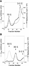
Fig. 2. Fractionation of the components of the in vitro translation system cross-linked to 32P-labeled [s4U]tmRNA. (A) Isolation of [s4U]tmRNA–70S ribosome complexes from the UV-irradiated in vitro translation system by centrifugation through the 14–40% sucrose gradient. (B) Separation of the [s4U]tmRNA–70S ribosome complexes into 30 and 50S ribosomal subunits by centrifugation through the 10–30% sucrose gradient. Dashed lines represent UV absorbance and closed squares trace the radioactivity profile. (C) Electrophoretic analysis of proteins cross-linked to [32P][s4U]tmRNA. The in vitro translation system containing [32P][s4U]tmRNA before irradiation (lane 1) and after irradiation (lane 2), as well as 70S (lane 3) and 3–5S (lane 4) fractions from the sucrose gradient (A), were treated by RNase T1 and subjected to Tricine–SDS–PAGE. The positions of protein S1 labeled by a fragment of tmRNA (solid arrow) and molecular weight markers are indicated.
None
Fig. 3. Cross-linking of 32P-labeled [s4U]tmRNA to wild-type and protein S1-depleted ribosomes. Aliquots of UV-irradiated [s4U]tmRNA (lane 1), complexes of [s4U]tmRNA and protein S1 (lanes 2 and 5), complexes of [s4U]tmRNA and wild-type 70S ribosomes (lanes 3 and 6), and complexes of [s4U]tmRNA and 70S ribosomes lacking protein S1 (lanes 4 and 7), as well as non-irradiated [s4U]tmRNA (Co), were digested with RNase T1 and subjected to Tricine–SDS–PAGE. Complexes indicated by a solid arrow were identified as [s4U]tmRNA–protein complexes by their susceptibility to proteinase K (lanes 5, 6 and 7). The positions of molecular weight markers are indicated.
None
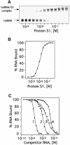
Fig. 4. Gel-shift analysis of binding between 32P-labeled tmRNA and protein S1His. (A) Titration of 3′-32P-labeled tmRNA (0.5 nM) with protein S1. Aliquots of binding mixtures were analyzed by electrophoresis on a 5% polyacrylamide gel (40:1) in TGE buffer. (B) Graphical representation of the PhosphorImager-derived data from the gel in (A). (C) Binding of [32P]tmRNA (1 nM) to protein S1 (100 nM) in the presence of unlabeled competitor RNAs: (1) tmRNA transcript; (2) poly(U); (3) tmRNAΔ90–299; and (4) E.coli tRNA.
None
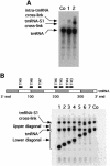
Fig. 5. Cross-linking tmRNA to ribosomal protein S1 and RNase H analysis of covalent tmRNA–protein S1 complexes. (A) The 3′-32P-labeled [s4U]tmRNA (lane 1) and non-covalent [s4U]tmRNA– protein S1 complexes (lane 2) were irradiated with UV light for10 min and analyzed on a 6% Tricine–SDS–polyacrylamide gel. (Co) Non-irradiated 3′-32P-labeled [s4U]tmRNA. UV irradiation yields internally cross-linked tmRNA, which, after cleaving with RNase Hin the presence of oligodeoxyribonucleotide TM3, co-migrates with non-irradiated [s4U]tmRNA (not shown). (B) Unpurified UV-irradiated 3′-32P-labeled [s4U]tmRNA–protein S1 complexes (Co) were digested by RNase H in the presence of oligodeoxyribonucleotides TM3 (lane 1), TM4 (lane 2), TM5 (lane 3), TM6 (lane 4), TM7 (lane 5), TM8 (lane 6) and TM9 (lane 7) as listed in Materials and methods, and separated on a 6% Tricine–SDS–polyacrylamide gel. The sites of hybridization of the oligodeoxyribonucleotides (solid bars) in tmRNA are illustrated in the diagram.
None
Fig. 6. Primer extension analysis of nucleotides in [s4U]tmRNA molecules that, upon irradiation with near-UV light, are cross-linked to a free protein S1 (A) and a 70S ribosome-bound protein S1 (B). U, C, G and A, sequencing lanes; 1, non-irradiated tmRNA; 2, UV-irradiated tmRNA; 3, tmRNA cross-linked to protein S1. The examples of primer extension reactions depicted were carried out in the presence of primers TM2 and TM6.
None
Fig. 7. RNase T1 protection analysis of tmRNA–S1 cross-links. (A) Covalent complexes formed by UV irradiation of 32P-internally labeled [s4U]tmRNA and free protein S1 were first digested with RNase T1, either in the absence (control reaction, Co) or presence of oligodeoxyribonucleotides xl-85 (lane 1), xl-240 (lane 2) and xl-3′-end (lane 3), and then analyzed by electrophoresis on a 6% polyacrylamide gel in Tricine–SDS buffer. Cross-linked segments of tmRNA that are protected by oligodeoxyribonucleotides xl-85 and xl-240 are indicated by a solid arrow. Other tmRNA segments protected only by protein S1 are indicated by an open arrow. (B) The same analysis carried out on cross-linked tmRNA–protein S1 complexes isolated from ribosomal particles using 5′-biotinylated oligodeoxyribonucleotides xl-105B (lane 1), xl-240B-1 (lane 2), xl-212B (lane 3) and xl-240B-2 (lane 4). The sites of hybridization of the oligodeoxyribonucleotides (solid bars) in tmRNA are illustrated in the diagram. Since the analyzed samples were purified on Ni2+-NTA–agarose prior to electrophoresis, cross-linked tmRNA fragments protected only by protein S1 are not present on the gel. The positions of molecular weight markers are indicated.
None
Fig. 8. Distribution of the sites of cross-linking to protein S1 in the secondary structure of the E.coli tmRNA. Squares and circles indicate the nucleotides of [s4U]tmRNA cross-linked to protein S1 on and off the ribosome, respectively. Letters ‘t’ and ‘m’ identify tRNA- and mRNA-like domains of tmRNA. Pseudoknots are labeled pk1–pk4. Solid arrows indicate sites of deletion that yielded tmRNAΔ90–299. Cross-links define the domain of tmRNA that is available for interactions with protein S1.

References

    1. Barends S., Wower,J. and Kraal,B. (2000) Kinetic parameters for tmRNA binding to alanyl-tRNA synthetase and elongation factor Tu from Escherichia coli. Biochemistry, 39, 2652–2658. - PubMed
    1. Barrera I., Schuppli,D., Sogo,J.M. and Weber H. (1993) Different mechanisms of recognition of bacteriophage Qβ plus and minus strand RNAs by Qβ replicase. J. Mol. Biol., 232, 512–521. - PubMed
    1. Bear D.G., Ng,R., Van Derveer,D., Johnson,N.P., Thomas,G., Schleich,T. and Noller,H.F. (1976) Alteration of polynucleotide secondary structure by ribosomal protein S1. Proc. Natl Acad. Sci. USA, 73, 1824–1828. - PMC - PubMed
    1. Berkower I., Leis,J. and Hurwitz,J. (1973) Isolation and characterization of an endonuclease from Escherichia coli specific for ribonucleic acid in ribonucleic acid–deoxyribonucleic acid hybrid structures. J. Biol. Chem., 248, 5914–5921. - PubMed
    1. Brimacombe R., Greuer,B., Gulle,H., Kosack,M., Mitchell,P., Osswald,M., Stade,K. and Stiege,W. (1990) New techniques for the analysis of intra-RNA and RNA–protein cross-linking data from ribosomes. In Spedding,G. (ed.), Ribosomes and Protein Synthesis. A Practical Approach. Oxford University Press, Oxford, UK, pp. 131–159.

Publication types

MeSH terms

LinkOut - more resources