Molecular characterization and diagnostic value of Taenia solium low-molecular-weight antigen genes
- PMID: 11101577
- PMCID: PMC87618
- DOI: 10.1128/JCM.38.12.4439-4444.2000
Molecular characterization and diagnostic value of Taenia solium low-molecular-weight antigen genes
Abstract
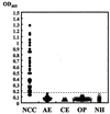
Neurocysticercosis (NCC) caused by infection with the larvae of Taenia solium is an important cause of neurological disease worldwide. In order to establish an enzyme-linked immunosorbent assay (ELISA) for this infection using recombinant proteins, we carried out molecular cloning and identified four candidates as diagnostic antigens (designated Ag1, Ag1V1, Ag2, and Ag2V1). Except for Ag2V1, these clones could encode a 7-kDa polypeptide, and Ag2V1 could encode a 10-kDa polypeptide. All of the clones were very similar. Except for Ag2V1, recombinant proteins were successfully expressed using an Escherichia coli expression system. Immunoblot analysis of NCC patient sera detected recombinant proteins, but because reactivity to recombinant Ag1 was too weak, Ag1 was not suitable as an immunodiagnostic antigen. So, Ag1V1 and Ag2 were chosen as ELISA antigens, and the Ag1V1/Ag2 chimeric protein was expressed. Of 49 serum samples from NCC patients confirmed to be seropositive by immunoblot analysis, 44 (89.7%) were positive by ELISA. No assays of serum samples from patients with other parasitic infections recognized the Ag1V1/Ag2 chimeric protein. The Ag1V1/Ag2 chimeric protein obtained in this study had a high value for differential immunodiagnosis.
Figures


References
-
- Baily G G, Mason P R, Trijssenar F E, Lyons N F. Serological diagnosis of neurocysticercosis: evaluation of ELISA tests using cyst fluid and other components of Taenia solium cysticerci as antigens. Trans R Soc Trop Med Hyg. 1988;82:295–299. - PubMed
-
- Brandt J R, Geerts S, De Deken R, Kumar V, Ceulemans F, Brijs L, Falla N. A monoclonal antibody-based ELISA for the detection of circulating excretory-secretory antigens in Taenia saginata cysticercosis. Int J Parasitol. 1992;22:471–477. - PubMed
-
- Chung J Y, Bahk Y Y, Huh S, Kang S Y, Kong Y, Cho S Y. A recombinant 10-kDa protein of Taenia solium metacestodes specific to active neurocysticercosis. J Infect Dis. 1999;180:1307–1315. - PubMed
-
- Craig P S, Rogan M T, Allan J C. Detection, screening and community epidemiology of taeniid cestode zoonoses: cystic echinococcosis, alveolar echinococcosis and neurocysticercosis. Adv Parasitol. 1996;38:169–250. - PubMed
-
- Fernandez V, Ferreira H B, Fernandez C, Zaha A, Nieto A. Molecular characterisation of a novel 8-kDa subunit of Echinococcus granulosus antigen B. Mol Biochem Parasitol. 1996;77:247–250. - PubMed
Publication types
MeSH terms
Substances
LinkOut - more resources
Full Text Sources
Miscellaneous

