

Acrophialophora fusispora brain abscess in a child with acute lymphoblastic leukemia: review of cases and taxonomy
- PMID: 11101597
- PMCID: PMC87638
- DOI: 10.1128/JCM.38.12.4569-4576.2000
Acrophialophora fusispora brain abscess in a child with acute lymphoblastic leukemia: review of cases and taxonomy
Abstract
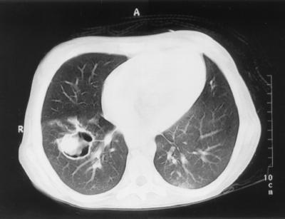
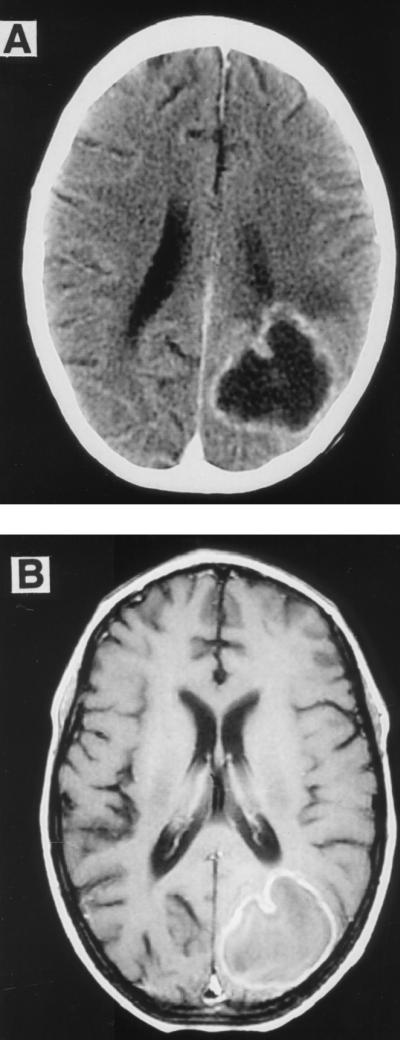
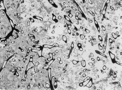
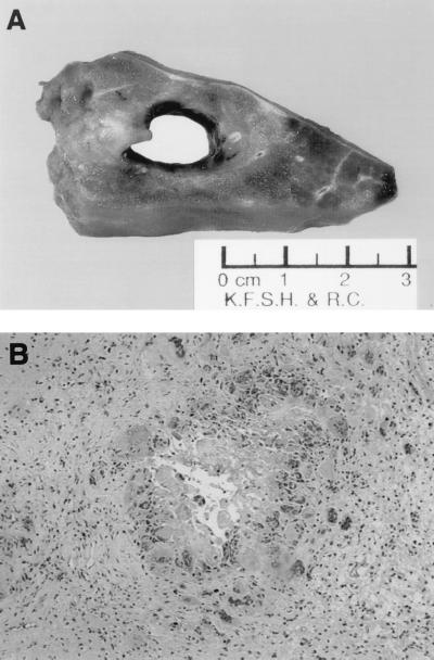
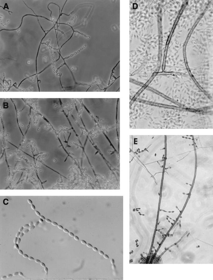
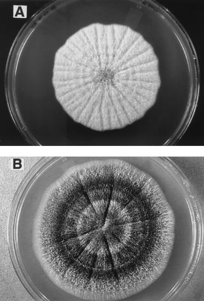
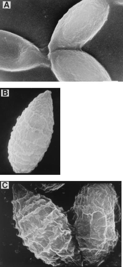
A 12-year-old girl with acute lymphoblastic leukemia was referred to King Faisal Specialist Hospital and Research Center. The diagnosis without central nervous system (CNS) involvement was confirmed on admission, and chemotherapy was initiated according to the Children Cancer Group (CCG) 1882 protocol for high-risk-group leukemia. During neutropenia amphotericin B (AMB) (1 mg/kg of body weight/day) was initiated for presumed fungal infection when a computed tomography (CT) scan of the chest revealed multiple nodular densities. After 3 weeks of AMB therapy, a follow-up chest CT revealed progression of the pulmonary nodules. The patient subsequently suffered a seizure, and a CT scan of the brain was consistent with infarction or hemorrhage. Because of progression of pulmonary lesions while receiving AMB, antifungal therapy was changed to liposomal AMB (LAMB) (6 mg/kg/day). Despite 26 days of LAMB, the patient continued to have intermittent fever, and CT and magnetic resonance imaging of the brain demonstrated findings consistent with a brain abscess. Aspiration of brain abscess was performed and the Gomori methenamine silver stain was positive for hyphal elements. Culture of this material grew Acrophialophora fusispora. Lung biopsy showed necrotizing fungal pneumonia with negative culture. The dosage of LAMB was increased, and itraconazole (ITRA) was added; subsequently LAMB was discontinued and therapy was continued with ITRA alone. The patient demonstrated clinical and radiological improvement. In vitro, the isolate was susceptible to low concentrations of AMB and ITRA. A. fusispora is a thermotolerant, fast-growing fungus with neurotropic potential. We report the first case of human infection involving the CNS. Acrophialophora resembles Paecilomyces but differs in having colonies that become dark and in the development of phialides along the sides or at the tips of echinulate brown conidiophores. Conidia are borne in long chains and are smooth or ornamented with fine-to-coarse echinulations, sometimes in spiral bands. The taxonomy of the genus Acrophialophora is reviewed, and Acrophialophora nainiana and Acrophialophora levis are considered as synonyms of A. fusispora.
Figures
References
-
- Anaissie E, Bodey G P, Kantarjian H, Ro J, Vartivarian S E, Hopfer R, Hoy J, Rolston K. New spectrum of fungal infections with cancer. Rev Infect Dis. 1989;11:369–378. - PubMed
-
- Barron G L. The genera of Hyphomycetes from soil. Baltimore, Md: Williams & Wilkins Co.; 1968.
-
- Brown A H S, Smith G. The genus Paecilomyces Bainer and its perfect stage Byssochlamys Wrestling. B Mycol Soc Trans. 1957;40:17–89.
-
- De Hoog G S, Guarro J. Atlas of clinical fungi. Baarn, The Netherlands: Centraalbureau voor Schimmelcutlures; 1995.
Publication types
MeSH terms
LinkOut - more resources
Full Text Sources
