Comparative Study
. 2000 Dec 1;529 Pt 2(Pt 2):483-92.
doi: 10.1111/j.1469-7793.2000.00483.x.

Accommodation to depolarizing and hyperpolarizing currents in cutaneous afferents of the human median and sural nerves

Affiliations
Comparative Study

Accommodation to depolarizing and hyperpolarizing currents in cutaneous afferents of the human median and sural nerves

C S Lin et al. J Physiol. .

Abstract

To determine whether accommodation to depolarizing and hyperpolarizing stimuli differs for cutaneous afferents in the median and sural nerves, studies were performed in normal human subjects using threshold electrotonus. The changes in threshold for compound sensory action potentials of 50 % of maximum were recorded when the nerves were subjected to long-lasting depolarizing and hyperpolarizing DC. The premise was that the threshold changes largely mirror the underlying electrotonic changes in membrane potential. The maximal threshold changes produced by depolarizing and hyperpolarizing currents were greater for median afferents, suggesting that the DC produced greater changes in membrane potential in these afferents. Median afferents underwent greater accommodation to depolarizing currents than sural afferents and a greater threshold undershoot at the end of the currents, suggesting greater activity of a slow K+ conductance. Median afferents also underwent greater accommodation to hyperpolarizing currents, suggesting greater inward rectification. These conductances are voltage dependent, and the differences in accommodation could be due to greater changes in membrane potential for the median nerve. The changes in threshold produced by long-lasting depolarizing and hyperpolarizing currents of graded intensity were therefore measured. When the threshold changes were matched for the two nerves, median afferents underwent 22.4 % more accommodation to depolarizing currents and 28.7 % more accommodation to hyperpolarizing currents. We conclude that there is greater expression of two internodally located conductances responsible for accommodation on median afferents. The biophysical differences identified in this study might contribute to the finding that sural afferents have a greater tendency to dysfunction than median afferents.


Figures
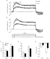
Figure 2
Figure 2. Threshold electrotonus to depolarizing currents
A, mean data (±s.e.m.) for eight subjects; •, median; ^, sural. The threshold changes due to 150 ms depolarizing currents of +20 and +40 % of the unconditioned threshold were measured. Median cutaneous afferents have a greater S1d, greater S2 and greater threshold undershoot on termination of the current than sural afferents. B shows that these findings were reproducible (means ±s.e.m. for 6 subjects). In C-E, data from A are plotted as histograms (means ±s.e.m.) for the two polarizing levels for the two nerves (□, median; ▪, sural). ‘Peak’ refers to the maximal threshold decrease and corresponds to the peak of S1d. S2 represents the difference between the peak threshold and the threshold at 150 ms. The ‘off response’ was measured 25–30 ms after the end of the DC pulse.
Figure 1
Figure 1. Threshold electrotonus in a single subject
Threshold electrotonus of median (•) and sural (^) cutaneous afferents from a single subject. The threshold required to produce a compound sensory action potential of 50 % of maximum was measured using test stimuli of 1.0 ms duration. The changes in threshold were produced by polarizing currents of 300 ms duration with intensities of +40, −40 and −80 % of the unconditioned threshold, as shown in the bottom panel. The symbols F, S1d, S1h, S2 and S3 are defined as shown for the responses of median afferents to 40 % depolarizing and 80 % hyperpolarizing currents (see also text). The three vertical lines were drawn at 25, 100 and 300 ms, i.e. at the peak of S1d, at the peak of S1h and at the end of the polarizing currents, respectively. d and h denote the depolarizing and hyperpolarizing direction, respectively.
Figure 3
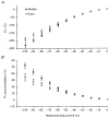
Figure 3. Polarizing level and the maximal threshold change to depolarizing currents
In A, F+S1d is the threshold measured 25 ms after the onset of depolarizing current. In B, S2 is the difference between thresholds measured 25 and 300 ms after the onset of the depolarizing current. Data are for the median nerve (•) and the sural nerve (^) for eight subjects (means ±s.e.m.).
Figure 4
Figure 4. Polarizing level and the maximal threshold change to hyperpolarizing currents
In A, S1h is the threshold measured 100 ms after the onset of hyperpolarizing current minus the fast (F) threshold change. In B, S3 is the difference between thresholds measured 100 and 300 ms after the onset of the hyperpolarizing current. Data represent means ±s.e.m. for the median nerve (•) and the sural nerve (^) for eight subjects.
Figure 5
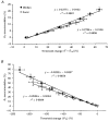
Figure 5. Relationship between accommodation and the driving threshold change
A, responses to depolarizing currents. F+S1d is the threshold measured 25 ms after the onset of the depolarizing current, presumably reflecting the change in membrane potential that drives S2. The relationship between the S2 accommodation and F+S1d is approximately linear for both nerves. The probability that the two lines have the same slope is 0.0134. B, responses to hyperpolarizing currents. S1h is the threshold change measured 100 ms after the onset of the hyperpolarizing current minus F, the fast phase, and presumably reflects the change in membrane potential that drives S3. The relationship between the S3 accommodation and S1h is approximately linear for both nerves. The probability that they have the same slope is < 0.001. •, median; ^, sural.

References

    1. Baker M, Bostock H. Depolarization changes the mechanism of accommodation in rat and human motor axons. The Journal of Physiology. 1989;411:545–561. - PMC - PubMed
    1. Baker M, Bostock H, Grafe P, Martius P. Function and distribution of three types of rectifying channel in rat spinal root myelinated axons. The Journal of Physiology. 1987;383:45–67. - PMC - PubMed
    1. Barrett EF, Barrett JN. Intracellular recording from vertebrate myelinated axons: mechanism of the depolarizing afterpotential. The Journal of Physiology. 1982;323:117–144. - PMC - PubMed
    1. Birch BD, Kocsis JD, Di Gregorio F, Bhisitkul RB, Waxman SG. A voltage- and time-dependent rectification in rat dorsal spinal root axons. Journal of Neurophysiology. 1991;66:719–728. - PubMed
    1. Bostock H. Mechanisms of accommodation and adaptation in myelinated axons. In: Waxman SG, Kocsis JD, Stys PK, editors. The Axon. New York: Oxford University Press; 1995. pp. 311–327.

Publication types

LinkOut - more resources