. 2000 Dec 1;20(23):8643-50.
doi: 10.1523/JNEUROSCI.20-23-08643.2000.

GABAC receptor sensitivity is modulated by interaction with MAP1B

Affiliations

GABAC receptor sensitivity is modulated by interaction with MAP1B

D Billups et al. J Neurosci. .

Abstract

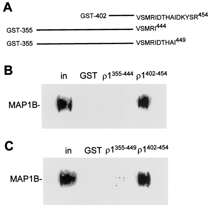
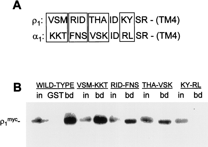
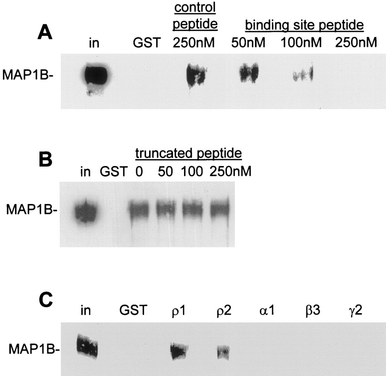
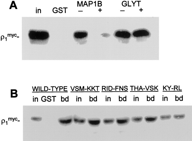
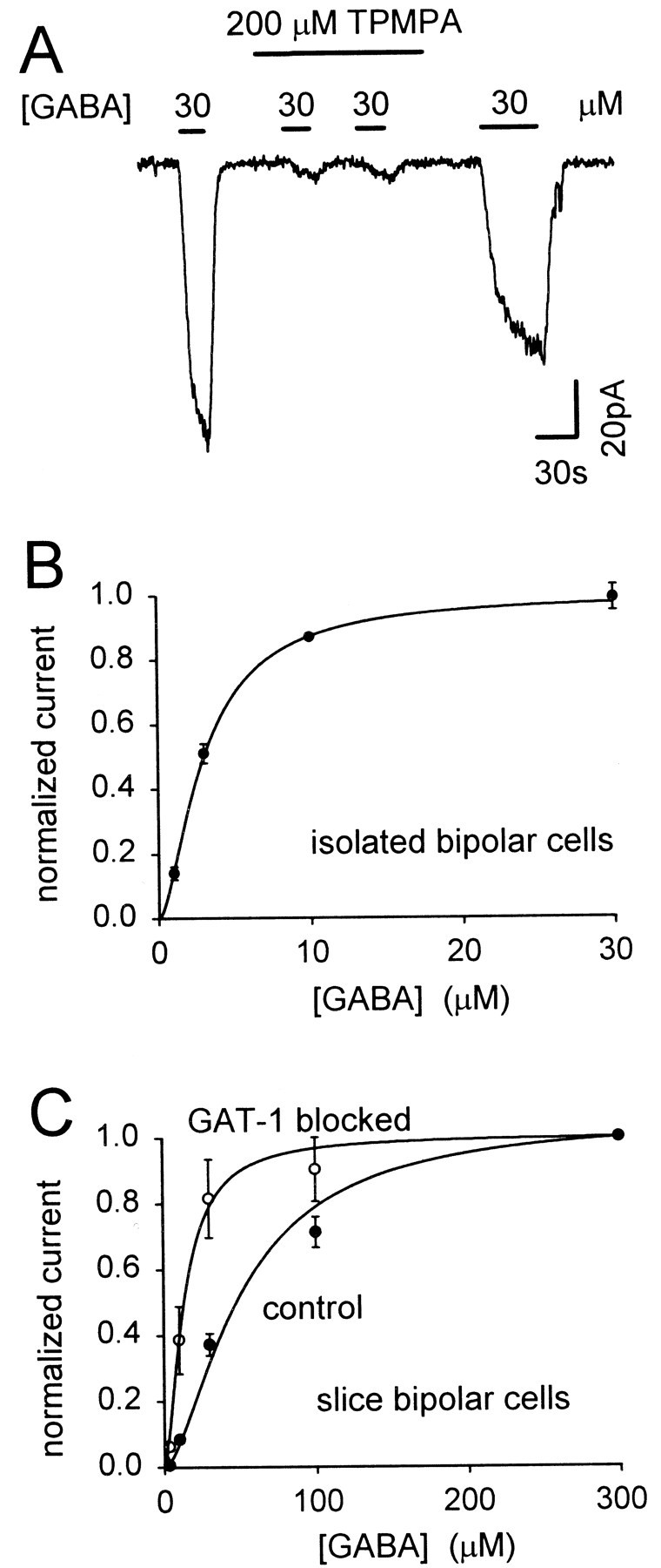
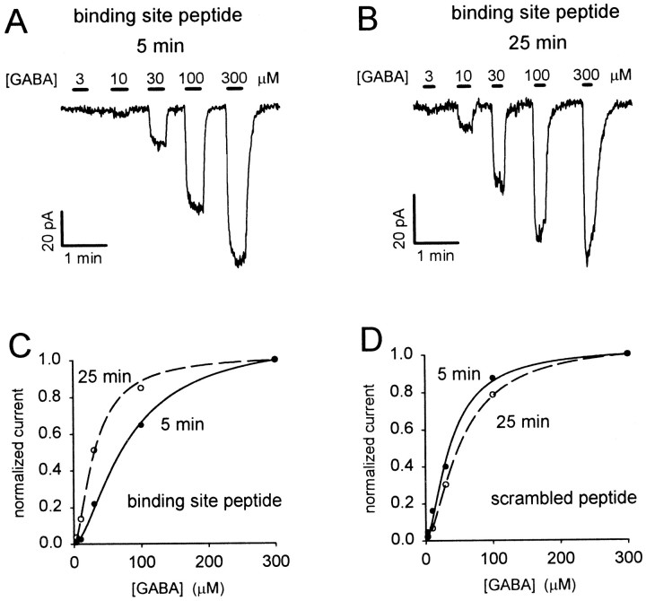
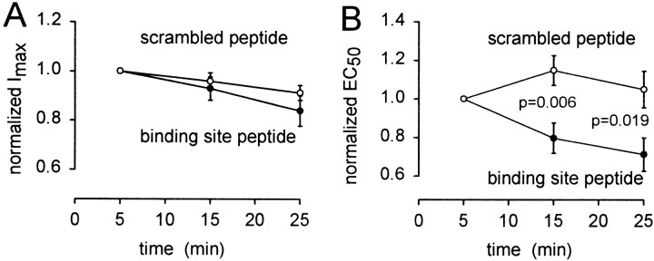
GABA(C) receptors contain rho subunits and mediate feedback inhibition from retinal amacrine cells to bipolar cells. We previously identified the cytoskeletal protein MAP1B as a rho1 subunit anchoring protein. Here, we analyze the structural basis and functional significance of the MAP1B-rho1 interaction. Twelve amino acids at the C terminus of the large intracellular loop of rho1 (and also rho2) are sufficient for interaction with MAP1B. Disruption of the MAP1B-rho interaction in bipolar cells in retinal slices decreased the EC(50) of their GABA(C) receptors, doubling the receptors' current at low GABA concentrations without affecting their maximum current at high concentrations. Thus, anchoring to the cytoskeleton lowers the sensitivity of GABA(C) receptors and provides a likely site for functional modulation of GABA(C) receptor-mediated inhibition.


Figures

Fig. 1.
Fig. 1.
MAP1B binds to the extreme C terminus of the ρ1 TM3–TM4 intracellular loop. Immobilized GST fusion proteins corresponding to C-terminal truncations of the ρ1 intracellular loop were incubated with retinal extract in pull-down assays. MAP1B binding was determined by Western blotting. A, Sequences of the GST fusions with the C-terminal half of ρ1 intracellular loop (ρ1402–454), with a 10 amino acid truncation (355–454) and a five amino acid truncation (355–449).B, Binding of MAP1B to the 10 amino acid truncation fusion protein, compared with binding to the C-terminal half of the intracellular loop ρ1402–454. Otherlanes show lack of binding to GST and the input to the assay. in represents the MAP1B present in 1% of the input. C, Same as B but for the five amino acid truncated fusion protein.
Fig. 2.
Fig. 2.
Identification of amino acid residues in the ρ1 intracellular loop important for MAP1B binding. Immobilized GST fusion protein corresponding to the ρ1-binding region of MAP1B was incubated with extracts of COS cells transfected with mutants of full-length ρ1myc in pull-down assays. Groups of residues in ρ1 were mutated to the equivalent sequence of the α1 subunit of GABAA receptors. A, Alignment of extreme C-terminal regions of intracellular TM3–TM4 loop of ρ1 and α1 subunits. Amino acid substitutions are shown in boxes; identical amino acids were not mutated. B, Binding of ρ1myc mutants to GST-MAP1B as determined by Western blotting. For each construct, in represents the ρ1myc present in 5% of the input, andbd represents protein bound to GST-MAP1B;GST shows lack of binding of ρ1mycto GST alone.
Fig. 3.
Fig. 3.
Competitive inhibition of MAP1B binding to ρ by peptides. The peptide containing the motif RINTHAIDKYSR (A) but not that containing THAIDKYSR (B) competes for MAP1B binding in retinal pull-down assays. A, Immobilized GST-ρ1 was incubated first with retinal extract, followed by 50–250 nm MAP1B binding site peptide or 250 nm scrambled control peptide. MAP1B bound to beads after this treatment was determined by Western blotting. B, Same as in A, but with truncated binding-site peptide. C, Pull-down assay from retinal lysate using immobilized GST alone or GST fusions of ρ1, ρ2, α1, β3, and γ2 subunit intracellular loops. For all panels, lane labeled GST shows no binding of MAP1B to GST alone, and in represents MAP1B present in 1% of input.
Fig. 4.
Fig. 4.
GLYT-1E/F and MAP1B interact with different regions of the ρ1 intracellular loop. A, Immobilized GST-GLYT-1E/F or GST-MAP1B were incubated first with extract of COS cells transfected with ρ1myc, followed by 250 nm of the MAP1B binding site peptide (+) containing RINTHAIDKYSR, or of the peptide containing a scrambled version (−) of the binding site. ρ1myc bound to beads after this treatment was determined by Western blotting. GST shows lack of binding of ρ1myc to GST alone, andin represents the ρ1myc present in 5% of the input. B, Immobilized GST-GLYT-1E/F was incubated with extracts of COS cells transfected with mutants of ρ1myc subunit in pull-down assays. Groups of residues in ρ1 were mutated to the equivalent sequence of α1 subunit of GABAA receptors (Fig. 2). For each construct,in represents the ρ1myc present in 5% of the input, and bd represents protein bound to GST-MAP1B; GST shows lack of binding of ρ1myc to GST alone.
Fig. 5.
Fig. 5.
GABAC receptor-evoked currents in retinal bipolar cells with no peptide included in the pipette solution.A, Membrane current of a cell in a slice, clamped to −60 mV, during repeated application of 30 μm GABA in the presence of 300 μm bicuculline to block GABAAreceptors. The GABAC receptor blocker TPMPA (200 μm) greatly reduces the response to GABA. This block was only slowly removed on washing out TPMPA. B, Dose–response curve for the application of GABA to isolated bipolar cells in the presence of 300 μm bicuculline (normalized to the response to 10 μm GABA in each cell and then rescaled to a saturating response of 1). Curve is a Hill equation (Eq.5) with n = 1.6 and a mean EC50 of 3 μm. Data from seven cells. C, Dose–response curves for five bipolar cells in slices, in the presence of 300 μm bicuculline and 4 mmCo2+/0 mm Ca2+, initially in normal conditions (control) and then in the presence of 100 μm SKF89976A (GAT-1 blocked). Hill equations through the data have n = 1.6 and an EC50 of 51 μm in control conditions (mean value was 51.0 ± 3.7 μm) and 13.4 μm with GAT-1 blocked (mean value was 13.4 ± 2.2 μm).
Fig. 6.
Fig. 6.
Effect of dialysis with MAP1B binding site peptide on the GABA dose–response curve in retinal bipolar cells.A, Specimen current responses to different GABA concentrations (in the presence of bicuculline) 5 min after going to whole-cell mode, with the binding site peptide in the pipette.B, Dose–response curve in the same cell asA, 25 min after starting whole-cell clamping. Responses in A and B have been scaled to be the same for 300 μm GABA, to compensate for a slight decline with time (Fig. 7A), and to facilitate comparison of the dose dependence of the responses. The relative responses to low doses of GABA are much larger in B than in A.C, Dose–response data from A andB, normalized to the current produced by 300 μm GABA, fitted with the Hill equation (Eq. 5) with a Hill coefficient of 1.6. At 5 min the EC50 is 79 μm; at 25 min it is 31 μm.d, Specimen dose–response data as in C, but from a cell clamped with the scrambled peptide in the pipette. Fitting the Hill equation (with a Hill coefficient of 1.6) gives an EC50 of 37 μm at 5 min and 53 μm at 25 min after starting whole-cell clamping.
Fig. 7.
Fig. 7.
Time-dependent changes in the properties of GABAC receptors induced by competitive removal of MAP1B binding. Maximum current (A) and EC50(B) derived from fitting the Hill equation (Eq.5) to dose–response data measured at different times after starting whole-cell clamping with pipette solution containing the MAP1B binding site peptide (●) or a scrambled version of it (○). Data from each cell were normalized to their values measured 5 min after starting whole-cell clamping, before averaging for these graphs. Data inA are not significantly different for the two peptides. The p values in B are from two-tailedt tests. Eight to thirteen cells contribute to each data point.

References

    1. Bonnert TP, McKernan RM, Farrar S, Le Bourdelles B, Heavens RP, Smith DW, Hewson L, Rigby MR, Sirinathsinghji DJS, Brown N, Wafford KA, Whiting PJ. θ, a novel γ-aminobutyric acid type A receptor. Proc Natl Acad Sci USA. 1999;96:9891–9896. - PMC - PubMed
    1. Borden LA, Dhar TGM, Smith KE, Weinshank RL, Branchek TA, Gluchowski C. Tiagabine, SK&F 89976-A, CI-966 and NNC-711 are selective for the cloned GABA transporter GAT-1. Eur J Pharmacol. 1994;269:219–224. - PubMed
    1. Calvo DJ, Miledi R. Activation of GABAρ1 receptors by glycine and β-alanine. NeuroReport. 1995;6:1118–1120. - PubMed
    1. Chang Y, Weiss DS. Channel opening locks agonist onto the GABAC receptor. Nat Neurosci. 1999;2:219–225. - PubMed
    1. Cutting GR, Lu L, O'Hara BF, Kasch LM, Montrose-Rafizadeh C, Donovan DM, Shimada S, Antonarakis SE, Guggino WB, Uhl GR, Kazazian HH., Jr Cloning of the γ-aminobutyric acid (GABA) ρ1 cDNA: a GABA receptor subunit highly expressed in the retina. Proc Natl Acad Sci USA. 1991;88:2673–2677. - PMC - PubMed

Publication types

MeSH terms

LinkOut - more resources