. 2000 Dec 1;20(23):8710-6.
doi: 10.1523/JNEUROSCI.20-23-08710.2000.

Dendritic and axonal targeting of type 5 metabotropic glutamate receptor is regulated by homer1 proteins and neuronal excitation

Affiliations

Dendritic and axonal targeting of type 5 metabotropic glutamate receptor is regulated by homer1 proteins and neuronal excitation

F Ango et al. J Neurosci. .

Abstract

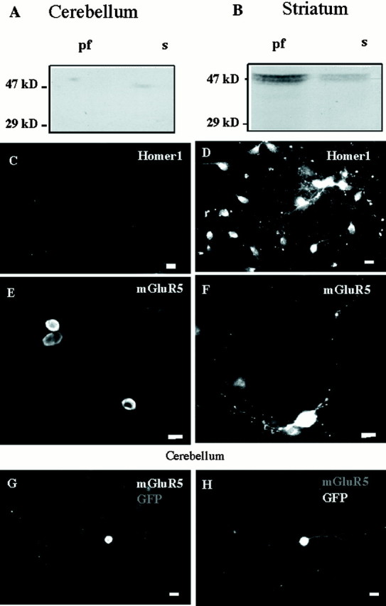
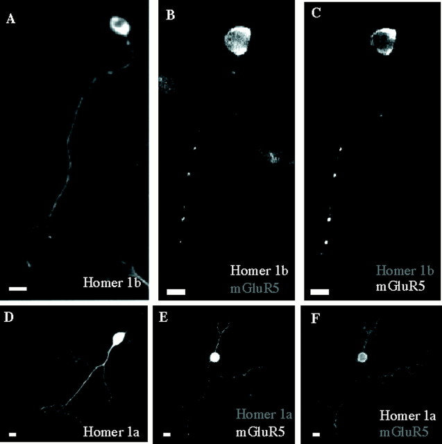
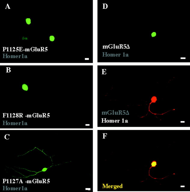
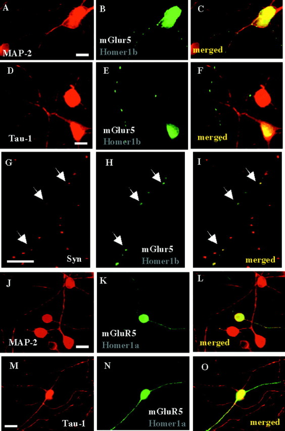
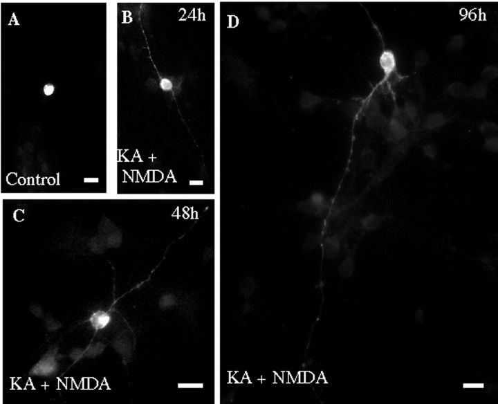
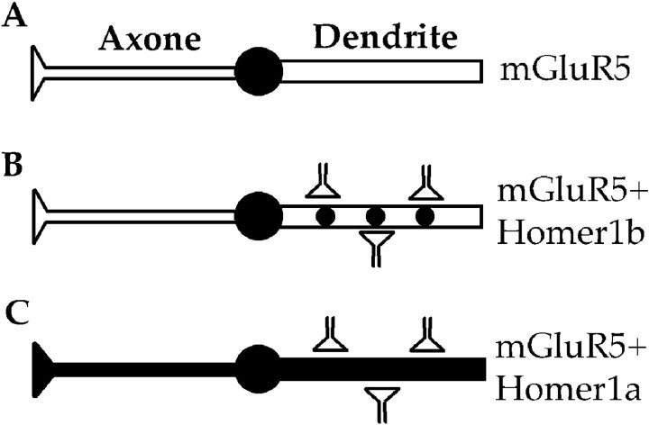
The physiological actions of neurotransmitter receptors are intimately linked to their proper neuronal compartment localization. Here we studied the effect of the metabotropic glutamate receptor (mGluR)-interacting proteins, Homer1a, b, and c, in the targeting of mGluR5 in neurons. We found that mGluR5 was exclusively localized in cell bodies when transfected alone in cultured cerebellar granule cells. In contrast, mGluR5 was found also in dendrites when coexpressed with Homer1b or Homer1c, and in both dendrites and axons when cotransfected with Homer1a. In dendrites, cotransfected mGluR5 and Homer1b/c formed clusters that colocalized with the synaptic marker synaptophysin. Interestingly when transfected alone, the Homer proteins were also translocated to neurites but did not form such clusters. Depolarization of the neurons with a mixture of ionotropic glutamate receptor agonists, NMDA and kainate, or potassium channel blockers, tetraethylammonium and 4-aminopyridine, induced transient expression of endogenous Homer1a and persistent neuritic localization of transfected mGluR5 even long after degradation of Homer1a. These results suggest that Homer1a/b/c proteins are involved in the targeting of mGluR5 to dendritic synaptic sites and/or axons and that this effect can be regulated by neuronal activity. Because the activity-dependent effect of endogenous Homer1a was also long-lasting, the axonal targeting of mGluR5 by this protein is likely to play an important role in synaptic plasticity.


Figures

Fig. 1.
Fig. 1.
Differential expression of native Homer1 and transfected mGluR5 proteins in striatal and cerebellar cultures.A, Western blot obtained from a 1-week-old cerebellar culture. The 29 kDa calibration mark is the apparent molecular weight of the Homer1a protein. Homer1b/c isoforms display apparent molecular weights of 47 kDa. pf, Particulate fraction;s, soluble fraction. Similar results were obtained in a second independent experiment. B, Same legend as inA, but from a 1-week-old striatal culture. Note the presence of Homer1b/c (47 kDa band). A andB were obtained from a same gel. C,Absence of Homer1 immunostaining in a 1-week-old cerebellar culture.D, Homer1a immunostaining in a 1-week-old striatal culture. E,F, HA–mGluR5 immunostaining with anti-HA antibody in transfected cerebellar granule cell (E) and striatal neuron (F). G,H, Same cerebellar granule cell cotransfected with HA–mGluR5 and GFP.G, HA immunostaining; H, GFP fluorescence. In this and all the following figures, scale bars represent 10 μm.
Fig. 2.
Fig. 2.
Neuritic localization of mGluR5 in cerebellar granule cells in the presence of Homer1a or Homer1b. A,Cerebellar neuron transfected with Myc–Homer1b and immunolabeled with anti-Myc antibody. B, C, Cerebellar neuron cotransfected with Myc–Homer1b and HA–mGluR5.B, Myc immunolabeling; C, HA immunolabeling in the same neuron as in B.D–F, Same legend as in A–C,respectively, but with epitope-tagged Homer1a.
Fig. 3.
Fig. 3.
Interaction between Homer1a and mGluR5 is required for neuritic localization of mGluR5 in cultured cerebellar granule cells. Cerebellar neurons were cotransfected with Homer1a (wild-type inA, B; Myc-tagged in C–E) and the indicated HA–mGluR5 mutants. A–D, HA immunolabeling. E, Myc immunolabeling in the same neuron as in D. F, Merged panelsD and E.
Fig. 4.
Fig. 4.
Axon–dendrite localization of mGluR5 in the presence of Homer1a or Homer1b, in cultured cerebellar granule cells.A–C, A 2-week-old cerebellar granule cell culture, previously cotransfected with Homer1b and HA–mGluR5, was co-immunolabeled with anti-MAP-2 (A) and anti-HA (B) antibodies. C, Merged panelsA and B. Note the presence of same HA- and MAP-2-immunoreactive neurite. D–F, Same legend as in A–C, but with anti-tau-1 antibody. Note the presence of HA-immunoreactive, but tau-1-negative neurite. G–I,Same culture as in A–F but co-immunolabeled with anti-synaptophysin (G) and anti-HA (H) antibodies. I, Merged panels G and H. Note the colocalization of HA–mGluR5 and synaptophysin clusters (arrows). Same size calibration as for A–F. Scale bars, 10 μm.J–L, M–O, Similar legend as in A–C, but in cerebellar neurons cotransfected with Homer 1a and HA–mGluR5.Arrows indicate HA, but not MAP-2 (J–L) or tau-1 (M–O) immunoreactive neurites. Note the presence of HA–mGluR5 in both MAP-2 (K, L) and tau-1 (N, O) immunoreactive neurites.
Fig. 5.
Fig. 5.
NMDA + kainate-induced transient expression of the immediate early gene product Homer1a in cultured cerebellar granule cells. A, Western blot obtained with an anti-Homer1 antibody, 12 hr after an NMDA + KA treatment. Note the presence of Homer1a protein (29 kDa band). B–E,Cerebellar cultures exposed to drug-free (B) or NMDA (100 μm) + kainate KA (100 μm)-containing culture medium (C–E) and labeled 12 hr (B–D) or 48 hr (E) later with the same anti-Homer1 antibody as in A. Note the absence of Homer1-immunoreactive neurons after 48 hr (E). Similar results were obtained with a TEA (20 mm) + 4-AP (20 mm) treatment.
Fig. 6.
Fig. 6.
Induction of Homer1a triggers localization of mGluR5 in the neurites of cultured cerebellar granule cells. Cerebellar cultures transfected with HA–mGluR5 were either exposed to drug-free (A) or NDMA + KA-containing culture medium (B–D) and immunolabeled with an anti-HA antibody. B–D were obtained 1, 2, and 4 d after the drug treatment, respectively. Note neuritic HA immunoreactivity in neurites of treated cultures (B–D).
Fig. 7.
Fig. 7.
Model for neuritic targeting of mGluR5 by Homer1 proteins. A, In the absence of Homer1, mGluR5 remains in the soma. B, Homer1b allows translocation and clustering of mGluR5 at dendritic synaptic sites. C, Induction of Homer1a expression triggers translocation of mGluR5 to both dendrites and axons.

Similar articles

Cited by

References

    1. Abe T, Sugihara H, Nawa H, Shigemoto R, Mizuno N, Nakanishi S. Molecular characterization of a novel metabotropic glutamate receptor mGluR5 coupled to inositol phosphate/Ca2+ signal transduction. J Biol Chem. 1992;267:13361–13368. - PubMed
    1. Aiba A, Kano M, Chen C, Stanton ME, Fox GD, Herrup K, Zwingman TA, Tonegawa S. Deficient cerebellar long-term depression and impaired motor learning in mGluR1 mutant mice. Cell. 1994;79:377–388. - PubMed
    1. Alagarsamy S, Marino MJ, Rouse ST, Gereau RW, Heinemann SF, Conn PJ. Activation of NMDA receptors reverses desensitization of mGluR5 in native and recombinant systems. Nat Neurosci. 1999;2:234–240. - PubMed
    1. Ango F, Albani-Torregrossa S, Joly C, Robbe D, Michel JM, Pin JP, Bockaert J, Fagni L. A simple method to transfer plasmid DNA into neuronal primary cultures: functional expression of the mGlu5 receptor in cerebellar granule cells. Neuropharmacology. 1999;38:793–803. - PubMed
    1. Anwyl R. Metabotropic glutamate receptors: electrophysiological properties and role in plasticity. Brain Res Rev. 1999;29:83–120. - PubMed

Publication types

MeSH terms

LinkOut - more resources