. 2000 Dec 1;20(23):8717-26.
doi: 10.1523/JNEUROSCI.20-23-08717.2000.

Presenilin-1 P264L knock-in mutation: differential effects on abeta production, amyloid deposition, and neuronal vulnerability

Affiliations

Presenilin-1 P264L knock-in mutation: differential effects on abeta production, amyloid deposition, and neuronal vulnerability

R Siman et al. J Neurosci. .

Abstract

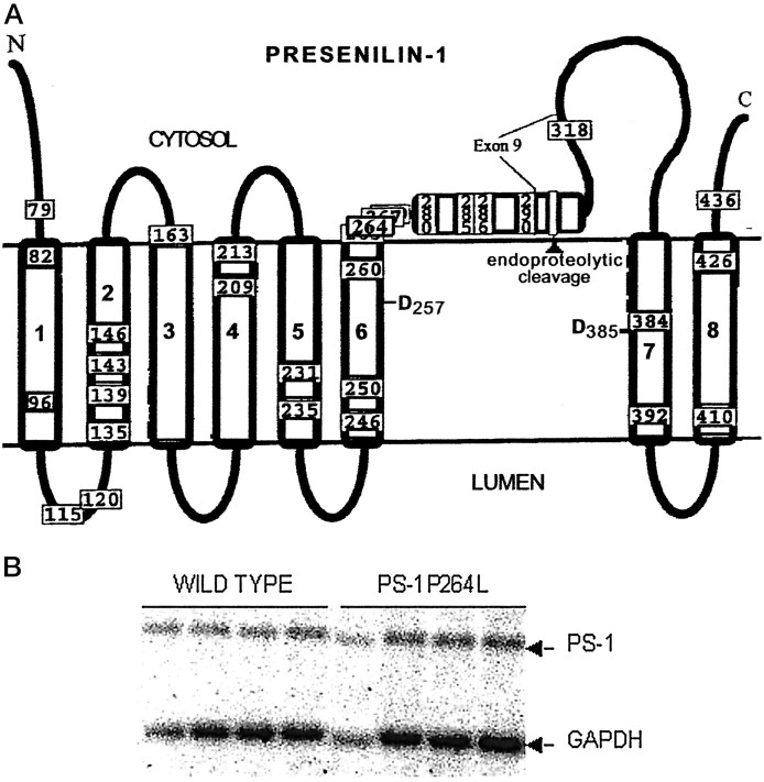
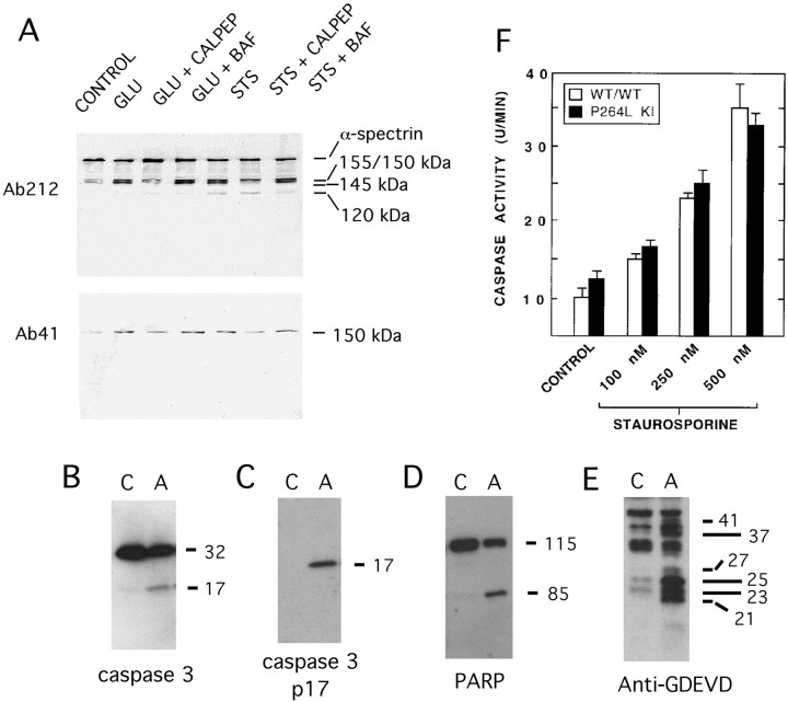
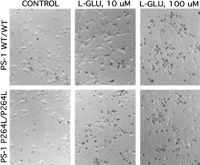
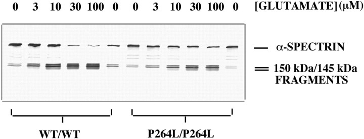
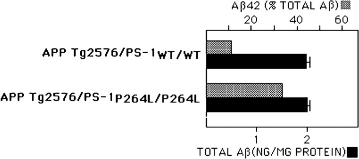
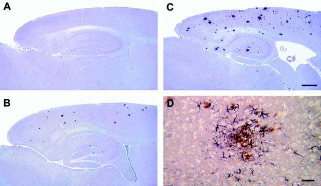
The pathogenic mechanism linking presenilin-1 (PS-1) gene mutations to familial Alzheimer's disease (FAD) is uncertain, but has been proposed to include increased neuronal sensitivity to degeneration and enhanced amyloidogenic processing of the beta-amyloid precursor protein (APP). We investigated this issue by using gene targeting with the Cre-lox system to introduce an FAD-linked P264L mutation into the endogenous mouse PS-1 gene, an approach that maintains normal regulatory controls over expression. Primary cortical neurons derived from PS-1 homozygous mutant knock-in mice exhibit basal neurodegeneration similar to their PS-1 wild-type counterparts. Staurosporine and Abeta1-42 induce apoptosis, and neither the dose dependence nor maximal extent of cell death is altered by the PS-1 knock-in mutation. Similarly, glutamate-induced neuronal necrosis is unaffected by the PS-1P264L mutation. The lack of effect of the PS-1P264L mutation is confirmed by measures of basal- and toxin-induced caspase and calpain activation, biochemical indices of apoptotic and necrotic signaling, respectively. To analyze the influence of the PS-1P264L knock-in mutation on APP processing and the development of AD-type neuropathology, we created mouse lines carrying mutations in both PS-1 and APP. In contrast to the lack of effect on neuronal vulnerability, cortical neurons cultured from PS-1P264L homozygous mutant mice secrete Abeta42 at an increased rate, whereas secretion of Abeta40 is reduced. Moreover, the PS-1 knock-in mutation selectively increases Abeta42 levels in the mouse brain and accelerates the onset of amyloid deposition and its attendant reactive gliosis, even as a single mutant allele. We conclude that expression of an FAD-linked mutant PS-1 at normal levels does not generally increase cortical neuronal sensitivity to degeneration. Instead, enhanced amyloidogenic processing of APP likely is critical to the pathogenesis of PS-1-linked FAD.


Figures

Fig. 1.
Fig. 1.
Location of P264L knock-in mutation introduced into PS-1 by gene-targeting. A, The eight transmembrane model for the topology of PS-1 is shown (Doan et al., 1996), along with mutations that have been linked to FAD (Hardy, 1997). Note that codon 264 is within a cluster site for FAD-linked mutations in the cytoplasmic loop region and is in proximity to one of the transmembrane aspartates (D257) implicated in regulating proteolytic function of γ-secretase. B, Northern analysis of PS-1 mRNA expression in brains of PS-1 wild-type and PS-1P264L/P264L mice. Levels of PS-1 and GAPDH mRNA were quantified by densitometry. Relative to GAPDH expression levels, knock-in of the P264L mutation did not alter expression of PS-1 mRNA.
Fig. 2.
Fig. 2.
Assessment of apoptosis in primary cortical neurons. Cultures were treated for 18 hr with either vehicle (A, B) or 600 nm STS (C, D). Shown inA and C are photomicrographs taken with modulation contrast optics, whereas B andD are fluorescence images of the same fields of chromatin staining with Hoechst 33342. Note the detectable but modest background level of apoptosis in vehicle-treated cultures, with some of the healthy neurons denoted by the arrows and an apoptotic profile indicated by the arrowhead. STS treatment caused abundant neuronal shrinkage, degeneration of processes, and condensation of chromatin indicative of apoptosis (arrowheads) in >70% of the neurons.
Fig. 3.
Fig. 3.
Cysteine protease activation during STS-induced apoptosis and l-glutamate-induced necrosis.A, Immunoblot detection of α-spectrin and its proteolytic fragments from cortical neurons treated withl-glutamate (100 μm) or STS (300 nm) in the presence or absence of the calpain inhibitor calpeptin (calpep, 50 μm) or the caspase inhibitor Boc-Asp(OMe)-FMK (BAF, 50 μm). An antibody to α-spectrin (Ab212) detects the intact ∼250 kDa polypeptide, as well as proteolytic fragments of ∼120–155 kDa. l-glutamate stimulates production of calpain-derived ∼150 and ∼145 kDa spectrin fragments, but not the ∼120 kDa caspase derivative. Calpain-mediated α-spectrin degradation is also detectable with Ab41, specific for a calpain-derived COOH-terminal ∼150 kDa fragment. B–E, Immunoblot detection of caspase-3 activation and caspase substrate degradation in apoptotic (A) but not control (C) cortical neurons. B, Procaspase-3 and the p17 subunit of activated caspase-3 detected with MAb46. C, The p17 subunit of activated caspase-3 detected with the neoepitope-specific antibody Ab206. D, Poly(ADP-ribose) polymerase (PARP; ∼115 kDa) and the caspase-derived ∼85 kDa fragment. E, Caspase substrate fragments containing the GDEVD caspase recognition motif, labeled with the neoepitope-specific antibody Ab127. F, Effect of PS-1 P264L/P264L on caspase activity in cortical neurons. Hydrolysis of the caspase substrate Ac-DEVD-AFC was quantified as described in Materials and Methods, and is represented as the time-dependent change in fluorescence per culture. Neither the basal level of caspase activity nor the STS-stimulated activity differed between the two PS-1 genotypes.
Fig. 4.
Fig. 4.
l-glutamate-induced necrosis is not altered by the PS-1 P264L/P264L knock-in mutation. Cortical neurons were treated for 24 hr with the indicated concentrations ofl-glutamate, then stained with Trypan blue to detect loss of plasma membrane integrity. Note that l-glutamate causes a dose-dependent neurotoxicity, and neither the basal- nor thel-glutamate-stimulated neuronal death was altered appreciably by the PS-1 point mutation. Hoffman modulation contrast optics, 200×.
Fig. 5.
Fig. 5.
Calpain-mediated spectrin degradation is not enhanced by PS-1 P264L/P264L. Western blot analysis of α-spectrin degradation using Ab212, after 4 hr l-glutamate treatment. Under basal conditions, α-spectrin exists predominantly as an ∼250 kDa polypeptide, with minor levels of ∼150/145 kDa fragments.l-glutamate causes a dose-dependent disappearance of intact α-spectrin and appearance of calpain-derived fragments of ∼150/145 kDa (Fig. 3). In the experiment shown, the basal- and glutamate-stimulated levels of spectrin degradation were modestly reduced by the PS-1 P264L mutation. Although this difference was not observed in two repeat experiments, in none of the experiments did the PS-1 point mutation enhance spectrin degradation.
Fig. 6.
Fig. 6.
PS-1P264L/P264L knock-in mutation elevates selectively Aβ42 level in the mouse brain. Whole brain extracts taken from 1-month-old mice were evaluated by ELISAs for Aβx-42 and Aβx-40 and normalized to total protein content. Although the total Aβ content does not differ between the two PS-1 genotypes, Aβ42 levels increased from 12% of the total Aβ to >30%. The difference is significant (p < 0.01).
Fig. 7.
Fig. 7.
PS-1P264L knock-in mutation accelerates amyloid deposition and reactive astrogliosis even as a single mutant allele. Sagittal sections immunostained for Aβ from 6-month-old APP695swe/PS-1wt/wt (A), 4-month-old APP695swe/PS-1 P264L/wt (B), and 6-month-old APP695swe/PS-1 P264L/wt (C, D). D is a higher magnification of double-labeling for Aβ (brown) and GFAP (purple) from parietal cortex. Scale bars:A–C, 500 μm; D, 50 μm.

Similar articles

Cited by

References

    1. Banker G, Goslin K. Culturing nerve cells. MIT; Cambridge, MA: 1998.
    1. Borchelt DR, Thinakaran G, Eckman CB, Lee MK, Davenport F, Ratovitski T, Prada CM, Kim G, Seekins S, Yager D, Slunt HH, Wang R, Seeger M, Levey AI, Gandy SE, Copeland NG, Jenkins NA, Price DL, Younkin SG, Sisodia SS. Familial Alzheimer's disease-linked presenilin 1 variants elevate Aβ1–42/1–40 ratio in vitro and in vivo. Neuron. 1996;17:1005–1013. - PubMed
    1. Borchelt DR, Ratovitski T, van Lare J, Lee MK, Gonzales V, Jenkins NA, Copeland NG, Price DL, Sisodia SS. Accelerated amyloid deposition in the brains of transgenic mice coexpressing mutant presenilin 1 and amyloid precursor proteins. Neuron. 1997;19:939–945. - PubMed
    1. Bursztajn S, DeSouza R, McPhie DL, Berman SA, Shioi J, Robakis NK, Neve RL. Overexpression in neurons of human presenilin-1 or a presenilin-1 familial Alzheimer disease mutant does not enhance apoptosis. J Neurosci. 1998;18:9790–9799. - PMC - PubMed
    1. Cai XD, Golde TE, Younkin SG. Release of excess amyloid β protein from a mutant amyloid β-protein precursor. Science. 1993;259:514–516. - PubMed

Publication types

MeSH terms