. 2000 Dec 1;20(23):8736-44.
doi: 10.1523/JNEUROSCI.20-23-08736.2000.

Localization and enhanced current density of the Kv4.2 potassium channel by interaction with the actin-binding protein filamin

Affiliations

Localization and enhanced current density of the Kv4.2 potassium channel by interaction with the actin-binding protein filamin

K Petrecca et al. J Neurosci. .

Abstract

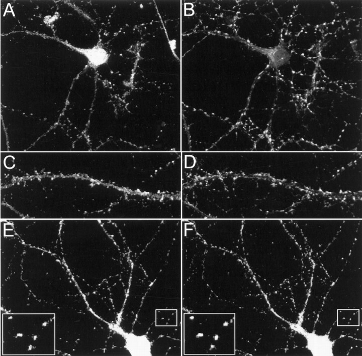
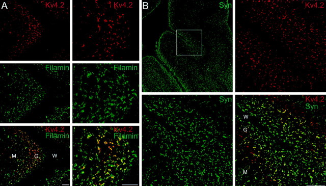
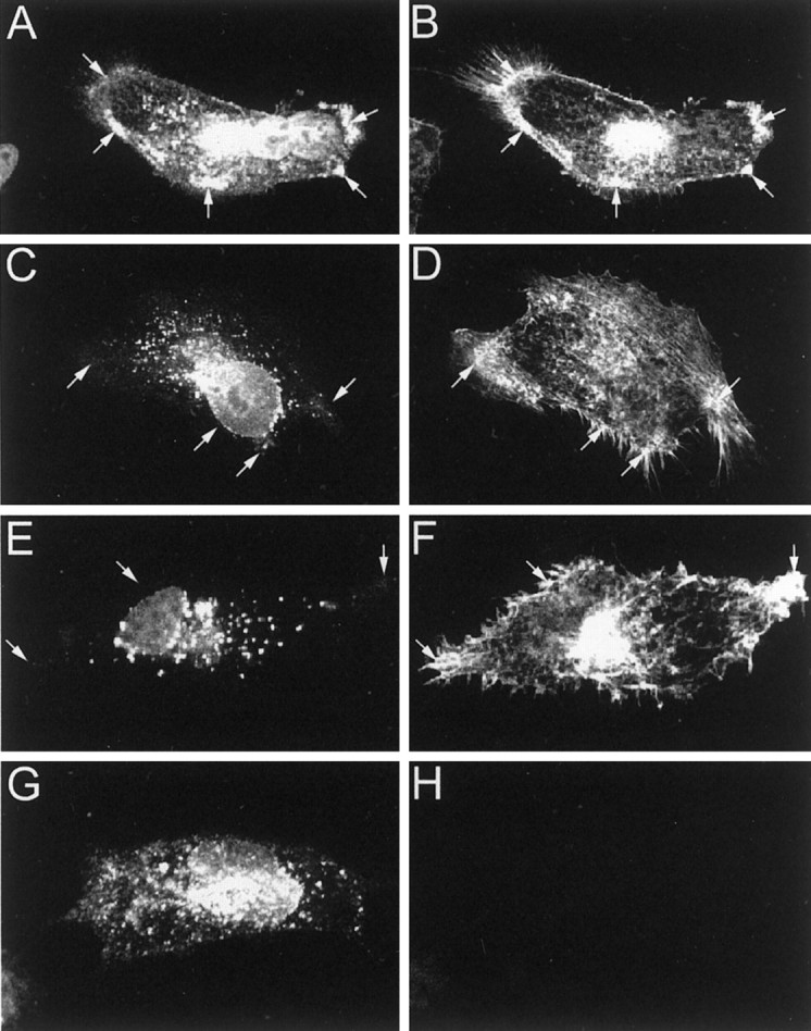
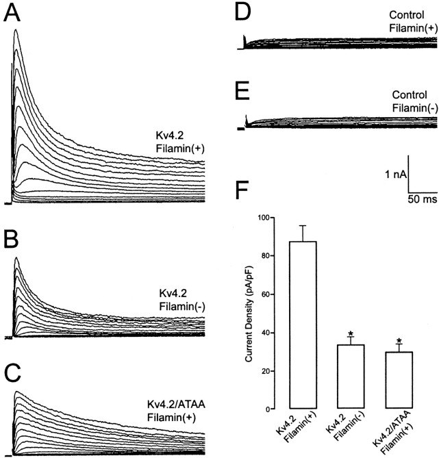
Kv4.2 potassium channels play a critical role in postsynaptic excitability. Immunocytochemical studies reveal a somatodendritic Kv4.2 expression pattern, with the channels concentrated mainly at dendritic spines. The molecular mechanism that underlies the localization of Kv4.2 to this subcellular region is unknown. We used the yeast two-hybrid system to identify the Kv4.2-associated proteins that are involved in channel localization. Here we demonstrate a direct interaction between Kv4.2 and the actin-binding protein, filamin. We show that Kv4.2 and filamin can be coimmunoprecipitated both in vitro and in brain and that Kv4.2 and filamin share an overlapping expression pattern in the cerebellum and cultured hippocampal neurons. To examine the functional consequences of this interaction, we expressed Kv4.2 in filamin(+) and filamin(-) cells and performed immunocytochemical and electrophysiological analyses. Our results indicate that Kv4.2 colocalizes with filamin at filopodial roots in filamin(+) cells but shows a nonspecific expression pattern in filamin(-) cells, with no localization to filopodial roots. Furthermore, the magnitude of whole-cell Kv4.2 current density is approximately 2.7-fold larger in filamin(+) cells as compared with these currents in filamin(-) cells. We propose that filamin may function as a scaffold protein in the postsynaptic density, mediating a direct link between Kv4.2 and the actin cytoskeleton, and that this interaction is essential for the generation of appropriate Kv4.2 current densities.


Figures

Fig. 1.
Fig. 1.
The domain structure of filamin and interaction with Kv4.2. A, Human cDNA clones isolated with a yeast two-hybrid screen, using the Kv4.2 C-terminal region (aa 395–630) as bait, are shown aligned below a schematic representation of the filamin domain structure. ABD, Actin binding domain;1–15, 16–23, and 24represent ∼96 aa repeats, each separated by hinge regions. Partial cDNAs from filaminA and filaminC genes were isolated. The numbers in parenthesesindicate the number of times each clone was isolated with the yeast two-hybrid screen. B, Sequence requirements in the Kv4.2 C-terminal region for interaction with filamin. FilaminC(aa 2172–2705), binding to Kv4.2C1 (aa 395–630), and deletion derivatives were assayed by HIS3/β-gal induction in the yeast two-hybrid system. Residues 601–604 are required for interaction with filamin; deletion and/or mutation of this region abolishes the interaction. Kv4.3, which contains the identical binding region, also interacts with filamin. The HERG C-terminal region (aa 864–1165) does bind filamin. The various bait fragments were tested for filamin binding by semiquantitative yeast two-hybrid interaction assays that were based on the degree of induction by the reporter genesHIS3 and β-gal. HIS3 activity was measured by the percentage of colonies growing on histidine-lacking medium as compared with the full-length Kv4.2 bait (Kv4.2C1): +++, >75%; ++, >50%; +, >25%. β-Gal activity was determined from the time that was taken for the colonies to turn blue in X-gal filter lift assays performed at room temperature: +++, <2 hr; ++, <3 hr; +, <4 hr; −, no significant activity.H6, Sixth transmembrane domain.
Fig. 2.
Fig. 2.
Coimmunoprecipitation in heterologous cells and direct binding of Kv4.2 and filamin. A, Extracts from COS7 cells singly or doubly transfected with HA-filamin and myc Kv4.2, myc-Kv4.2/600, myc-Kv4.2/ATAA, or myc-HERG were immunoprecipitated with anti-HA antibodies. The immunoprecipitates were immunoblotted with anti-myc (top panel) and anti-HA antibodies (bottom panel). B, Extracts from Kv4.2-transfected filamin+ and filamin cells were immunoprecipitated with anti-Kv4.2 antibodies and immunoblotted with anti-filamin (top panel) and anti-Kv4.2 (bottom panel) antibodies. C, Filter overlay assay showing direct in vitro binding of [35S]filamin to Kv4.2. GlutathioneS-transferase (GST) and GST-Kv4.2 (aa 417–630) fusion proteins were prepared as crude bacterial lysates and were purified with glutathione-Sepharose beads. Protein (5 μg) was resolved by SDS-PAGE and transferred to a PVDF membrane. Top panel, Renatured membrane overlaid with [35S]filamin showing specific binding to GST-Kv4.2. Bottom panel, Ponceau S-stained membrane showing the position and similar abundance of proteins in each lane.D, Filter overlay assay showing direct in vitro binding of HA-filamin to in vitro-translated Kv4.2, but not to Kv4.2/600 nor Kv4.2/ATAA. In all, 15 μl of in vitro-translated [35S]Kv4.2, [35S] Kv4.2/600, and [35S]Kv4.2/ATAA was resolved by SDS-PAGE and transferred to a PVDF membrane. Top panel, Renatured membrane overlaid with in vitro-translated HA-filamin and immunoblotted with anti-HA showing specific binding to Kv4.2, but not to Kv4.2/600 nor Kv4.2/ATAA. Bottom panel, The identical blot was stripped and exposed to autoradiography, showing the position and similar abundance of the in vitro-translated protein products in each lane.
Fig. 3.
Fig. 3.
Biochemical association of Kv4.2 and filamin in rat brain. Shown is the coimmunoprecipitation of Kv4.2 and filamin from cerebellum. Membrane fractions of cerebellar homogenate were solubilized and immunoprecipitated as indicated. A, The immunoblot shows that two different anti-filamin antibodies (lane 1, Sigma; lane 2, Serotec) specifically precipitate Kv4.2, as visualized by blotting with an anti-Kv4.2 antibody. Immunoprecipitation with control IgG (lane 5) does not pull down Kv4.2, demonstrating the specificity of the immunoprecipitation and competition of the anti-Kv4.2 antibody; the immunogenic peptide completely blocked the labeling of Kv4.2 (lane 4). The Input lane was loaded with 5% of the extract used for immunoprecipitation (lane 3). B, The immunoblot shows that two different anti-filamin antibodies (lane 2, Sigma;lane 3, Serotec) do not pull down HERG, as visualized by blotting with an anti-HERG antibody. The Input lane was loaded with 5% of the extract that was used for immunoprecipitation (lane 1).
Fig. 4.
Fig. 4.
Colocalization of Kv4.2 and filamin in cultured hippocampal neurons. Cultured hippocampal neurons were double-immunolabeled by using anti-Kv4.2 (A, C, E), anti-synaptophysin (B, D), and anti-filamin (F) antibodies. A–D, Double immunolabeling of endogenous Kv4.2 and synaptophysin shows that Kv4.2 is distributed in a punctate pattern along dendrites, matching closely with that of synaptophysin. E, F, Double immunolabeling of endogenous Kv4.2 and filamin shows that Kv4.2 and filamin colocalize in a punctate pattern along the dendrites. Large insets in E and F represent high magnification view of small insets in eachpanel, respectively.
Fig. 5.
Fig. 5.
Colocalization of Kv4.2 and filamin in cerebellum. Fresh-frozen 20-μm-thick cryosections of adult rat cerebellum were double-immunolabeled with anti-Kv4.2 (Cy3) and anti-filamin (Oregon green) antibodies (A) and anti-Kv4.2 (Cy3) and anti-synaptophysin (Oregon green) antibodies (B).A, Low-magnification (left) and high-magnification (right) images show that Kv4.2 and filamin colocalize in the cerebellar granule cell layer.B, Low- and high-magnification images (inset in top left panel indicates the region from which high-magnification images were obtained) show that the Kv4.2 and synaptophysin distribution patterns overlap.G, Granule cell layer; M, molecular layer; W, white matter. Scale bars: A, 50 μm; B, 100 μm.
Fig. 6.
Fig. 6.
Colocalization and accumulation of Kv4.2 and filamin at filopodial roots in filamin+ heterologous cells. Filamin+ M2 cells were transfected with myc-Kv4.2 (A, B), myc-Kv4.2/600 (C, D), and myc-Kv4.2/ATAA (E, F). Filamin M2 cells were transfected with myc-Kv4.2 (G, H). Cells were double-immunolabeled with anti-myc (A, C, E, G) and anti-filamin (B, D, F, H) antibodies. A, B, Kv4.2 exhibits a discrete subcellular distribution, colocalizing with filamin at filopodial roots in filamin+ M2 cells. C, D, Deletion of the C-terminal 30 aa of Kv4.2, including the filamin-binding site (Kv4.2/600), and substitution of the prolines with alanines within the filamin-binding site of Kv4.2 (Kv4.2/ATAA;E, F) results in a loss of Kv4.2 colocalization with filamin and a resulting nonspecific distribution with a marked absence at filopodial roots in filamin+ M2 cells.G, H, Kv4.2 exhibits a nonspecific distribution in filamin M2 cells. Arrows indicate Kv4.2 and filamin localization at filopodial roots.
Fig. 7.
Fig. 7.
Effect of the Kv4.2–filamin association on whole-cell Kv4.2 current density. Large, transient whole-cell currents were induced in filamin+ cells transfected with Kv4.2 (A) by 500 msec depolarizing steps from a −80 mV holding potential to potentials between −70 and 70 mV in 10 mV steps that were imposed at 10 sec intervals. Recordings from filamin cells transfected with Kv4.2 (B) and filamin+ cells transfected with Kv4.2/ATAA (C) revealed a smaller transient outward current. In untransfected filamin+ (D) or filamin (E) cells this transient current was not observed. Instead, a delayed rectifier-like endogenous current was recorded. Similar endogenous currents were revealed in filamin+ and filamin cells transfected with Kv4.2 by imposing the depolarizing step protocol from a −20 mV holding potential (data not shown). The differences in the magnitude of the transient outward current as measured from the initial transient outward peak to the current level at the end of the 500 msec depolarizing steps to +30 mV were found to be 2.6-fold greater in Kv4.2-transfected filamin+ cells (n = 22) than in filamin cells (n = 15; *p < 0.001) and 2.8-fold greater in Kv4.2-transfected filamin+ cells than in Kv4.2/ATAA-transfected filamin+ cells (n = 15; *p < 0.001). This difference is reflected in the bar graph (F).

Similar articles

Cited by

References

    1. Alonso G, Widmer H. Clustering of Kv4.2 potassium channels in postsynaptic membrane of rat supraoptic neurons: an ultrastructural study. Neuroscience. 1997;77:617–621. - PubMed
    1. Arnold DB, Clapham DE. Molecular determinants for subcellular localization of PSD-95 with an interacting K+ channel. Neuron. 1999;23:149–157. - PubMed
    1. Barry DM, Trimmer JS, Merlie JP, Nerbonne JM. Differential expression of voltage-gated K+ channel subunits in adult rat heart. Circ Res. 1995;77:361–369. - PubMed
    1. Burridge K, Chrzanowska-Wodnicka M. Focal adhesions, contractility, and signaling. Annu Rev Cell Dev Biol. 1996;12:463–518. - PubMed
    1. Colledge M, Froehner SC. To muster and cluster: anchoring neurotransmitter receptors and synapses. Proc Natl Acad Sci USA. 1998;95:3341–3343. - PMC - PubMed

Publication types

MeSH terms