

Differential postnatal development of catecholamine and serotonin inputs to identified neurons in prefrontal cortex of rhesus monkey
- PMID: 11102486
- PMCID: PMC6773090
- DOI: 10.1523/JNEUROSCI.20-23-08780.2000
Differential postnatal development of catecholamine and serotonin inputs to identified neurons in prefrontal cortex of rhesus monkey
Abstract
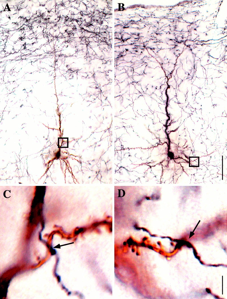
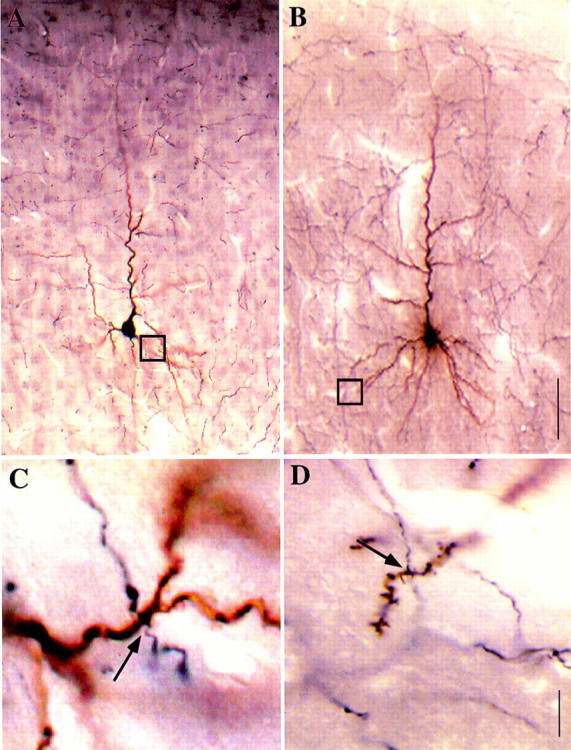
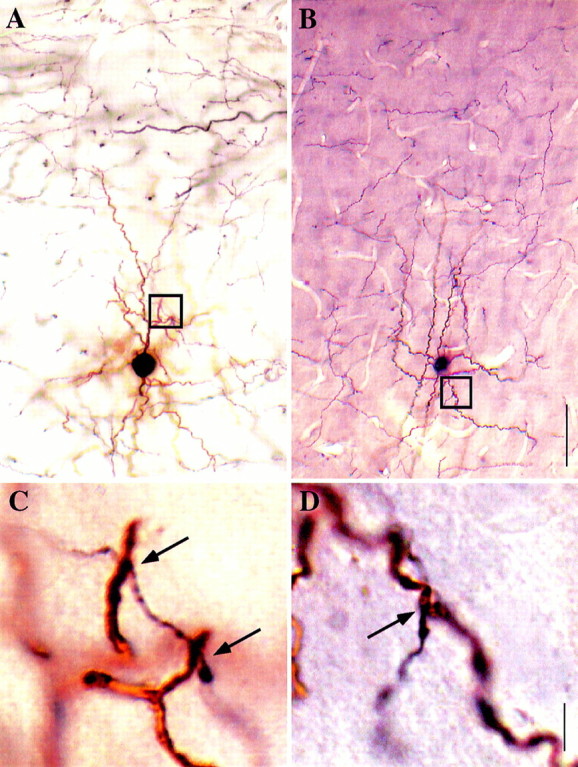
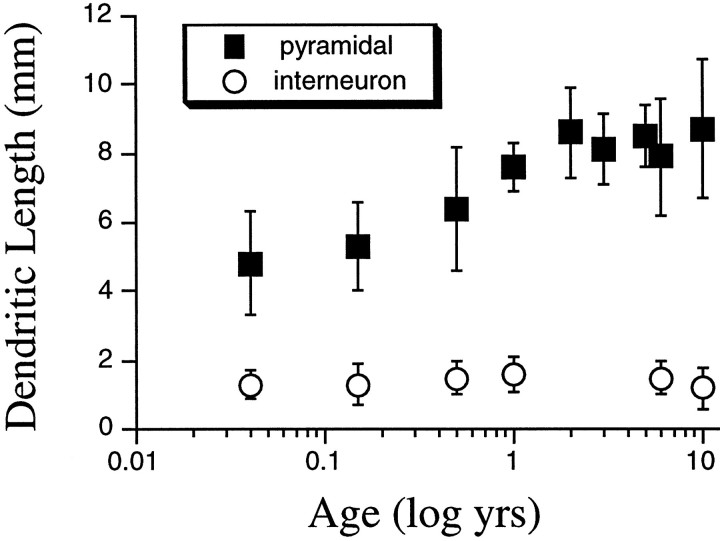
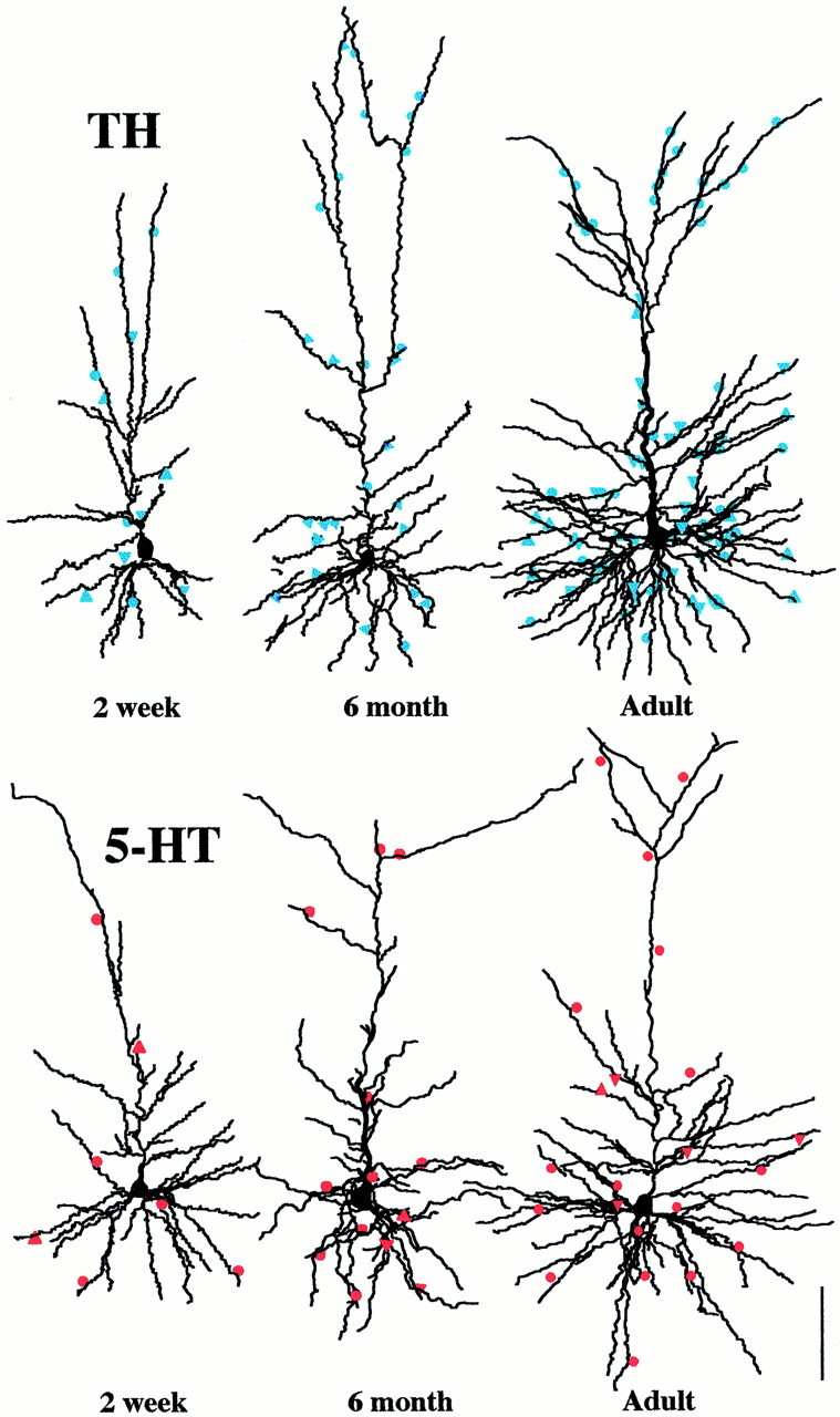
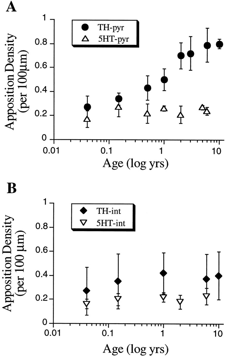
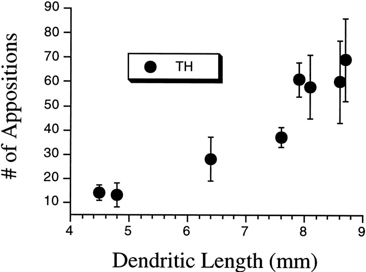
The monoaminergic innervation of cerebral cortex has long been implicated in its development. Methods now exist to examine catecholamine and serotonin inputs to identified neurons in the cerebral cortex. We have quantified such inputs on pyramidal and nonpyramidal cells in prefrontal cortex of rhesus monkeys ranging in age from 2 weeks to 10 years. Individual layer III neurons were filled with Lucifer yellow and double-immunostained with axons containing either tyrosine hydroxylase (TH) or 5-hydroxytryptamine (5-HT). The filled cells were reconstructed, and putative appositions between the axons and dendritic spines and shafts were quantified at high magnification using light microscopy. The density of catecholamine appositions on pyramidal neurons matures slowly, reaching only half the adult level by 6 months of age and thereafter rising gradually to adult levels by 2 years of age. By contrast, the density of serotonin appositions on pyramidal cells reaches the adult level before the second week after birth. The average adult pyramidal neuron in layer III of area 9m receives three times stronger input from catecholaminergic than from serotoninergic axons. The overall density of both inputs to interneurons does not appear to change during postnatal development. Selective changes in the TH innervation of pyramidal cells against a backdrop of constant TH innervation of interneurons suggest that the balance between excitation and inhibition may change developmentally in the prefrontal cortex. By contrast, 5-HT innervation of both types of neurons remains relatively constant over the age range studied.
Figures
Similar articles
-
Quantitative three-dimensional analysis of the catecholaminergic innervation of identified neurons in the macaque prefrontal cortex.J Neurosci. 1997 Oct 1;17(19):7450-61. doi: 10.1523/JNEUROSCI.17-19-07450.1997. J Neurosci. 1997. PMID: 9295391 Free PMC article.
-
Synchronous development of pyramidal neuron dendritic spines and parvalbumin-immunoreactive chandelier neuron axon terminals in layer III of monkey prefrontal cortex.Neuroscience. 1995 Jul;67(1):7-22. doi: 10.1016/0306-4522(95)00051-j. Neuroscience. 1995. PMID: 7477911
-
Serotonergic regulation of neuronal excitability in the prefrontal cortex.Neuropharmacology. 2011 Sep;61(3):382-6. doi: 10.1016/j.neuropharm.2011.01.015. Epub 2011 Jan 18. Neuropharmacology. 2011. PMID: 21251917 Free PMC article. Review.
-
Postnatal maturation of the dopaminergic innervation of monkey prefrontal and motor cortices: a tyrosine hydroxylase immunohistochemical analysis.J Comp Neurol. 1995 Jul 31;358(3):383-400. doi: 10.1002/cne.903580306. J Comp Neurol. 1995. PMID: 7560293
-
Modification of dendritic development.Prog Brain Res. 2002;136:135-43. doi: 10.1016/s0079-6123(02)36013-8. Prog Brain Res. 2002. PMID: 12143377 Review.
Cited by
-
Early Adolescence is a Critical Period for the Maturation of Inhibitory Behavior.Cereb Cortex. 2019 Aug 14;29(9):3676-3686. doi: 10.1093/cercor/bhy247. Cereb Cortex. 2019. PMID: 30295713 Free PMC article.
-
Development of prefrontal cortex.Neuropsychopharmacology. 2022 Jan;47(1):41-57. doi: 10.1038/s41386-021-01137-9. Epub 2021 Oct 13. Neuropsychopharmacology. 2022. PMID: 34645980 Free PMC article. Review.
-
Impacts of stress and sex hormones on dopamine neurotransmission in the adolescent brain.Psychopharmacology (Berl). 2014 Apr;231(8):1581-99. doi: 10.1007/s00213-013-3415-z. Epub 2014 Jan 31. Psychopharmacology (Berl). 2014. PMID: 24481565 Free PMC article. Review.
-
Norepinephrine versus dopamine and their interaction in modulating synaptic function in the prefrontal cortex.Brain Res. 2016 Jun 15;1641(Pt B):217-33. doi: 10.1016/j.brainres.2016.01.005. Epub 2016 Jan 11. Brain Res. 2016. PMID: 26790349 Free PMC article. Review.
-
Application of Research Domain Criteria to childhood and adolescent impulsive and addictive disorders: Implications for treatment.Clin Psychol Rev. 2018 Aug;64:41-56. doi: 10.1016/j.cpr.2016.11.003. Epub 2016 Nov 9. Clin Psychol Rev. 2018. PMID: 27876165 Free PMC article. Review.
References
-
- Aghajanian GK, Marek GJ. Serotonin induces excitatory postsynaptic potentials in apical dendrites of neocortical pyramidal cells. Neuropharmacology. 1997;36:589–599. - PubMed
-
- Akil M, Lewis DA. The dopaminergic innervation of monkey entorhinal cortex. Cereb Cortex. 1993;3:533–550. - PubMed
-
- Anderson SA, Classey JD, Conde F, Lund JS, Lewis DA. Synchronous development of pyramidal neuron dendritic spines and parvalbumin-immunoreactive chandelier neuron axon terminals in layer III of monkey prefrontal cortex. Neuroscience. 1995;67:7–22. - PubMed
-
- Aura J, Riekkinen PJ. Blockade of NMDA receptors located at the dorsomedial prefrontal cortex impairs spatial working memory in rats. NeuroReport. 1999;10:243–248. - PubMed
MeSH terms
Substances
LinkOut - more resources
Full Text Sources