. 2000 Dec 1;20(23):8831-7.
doi: 10.1523/JNEUROSCI.20-23-08831.2000.

NMDA and glutamate evoke excitotoxicity at distinct cellular locations in rat cortical neurons in vitro

Affiliations

NMDA and glutamate evoke excitotoxicity at distinct cellular locations in rat cortical neurons in vitro

J D Sinor et al. J Neurosci. .

Abstract

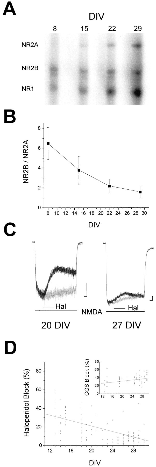
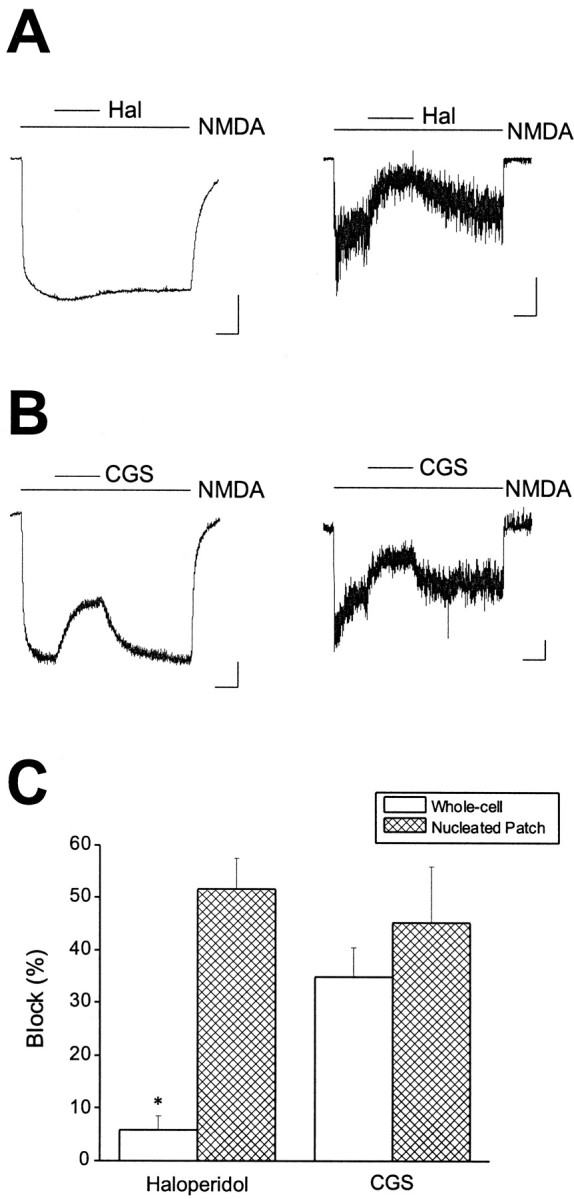
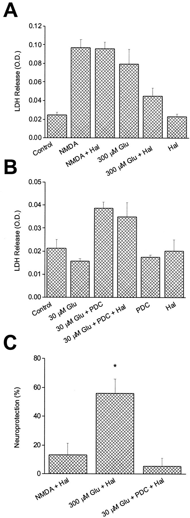
The development of cortical neurons in vivo and in vitro is accompanied by alterations in NMDA receptor subunit expression and concomitant modifications in the pharmacological profile of NMDA-activated ionic currents. For example, we observed that with decreasing NR2B/NR2A subunit expression ratio, the block of NMDA receptor-mediated whole-cell responses by the NR2B-selective antagonist haloperidol was also decreased. In mature cultures (>22 d in vitro), however, NMDA responses obtained from excised nucleated macropatches, which comprised a large portion of the soma, remained strongly antagonized by haloperidol. These results suggest that in more mature neurons NR1/NR2B receptors appear to be preferentially expressed in the cell body. As predicted from the whole-cell recording pharmacological profile, NMDA-induced toxicity was largely unaffected by haloperidol in mature cultures. However, haloperidol effectively blocked glutamate toxicity in the same cultures, suggesting that the neurotoxic actions of this amino acid were mostly due to the activation of somatic NMDA receptors. In experiments in which the potency of glutamate toxicity was increased by the transport inhibitor l-trans-pyrrolidine-2,4-dicarboxylic acid, the neuroprotective effects of haloperidol were significantly diminished. This was likely because of the fact that glutamate, now toxic at much lower concentrations, was able to reach and activate dendritic receptors under these conditions. These results strongly argue that exogenous glutamate and NMDA normally induce excitotoxicity at distinct cellular locations in mature mixed neuronal cultures and that NR1/NR2B receptors remain an important component in the expression of glutamate, but not NMDA-induced excitotoxicity.


Figures

Fig. 1.
Fig. 1.
Onset of NMDA receptor subunit expression in rat cortex in vitro. A, Representative gel from an RNase protection assay demonstrating the time course of NMDA receptor subunit expression in cortical neurons grown in astrocyte-rich cultures for up to 4 weeks. The expression of NMDA receptor subunits was determined by using 32P-lableled probes specific for NR1, NR2A, and NR2B.B, Quantification of the ratio of NR2B to NR2A band densities during the maturation of the cultures. Values represent the mean ± SEM of three separate experiments performed on three independent cultures. The decrease in band density ratio during development was statistically significant (p< 0.05; ANOVA). C, Whole-cell voltage-clamped (−60 mV) currents obtained from cortical neurons in astrocyte-rich cultures at two different developmental stages invitro. Currents were recorded during fast application of 30 μm NMDA in the absence or presence of 10 μm haloperidol (Hal). Thelinesbelow the traces indicate drug applications. Calibration: 150 pA, 1 sec. D, Responses such as those shown in C were used to measure the steady-state block by haloperidol of NMDA-elicited currents at various developmental time points. The measurements of current inhibition were corrected for the desensitization observed when agonist was applied alone (C, light traces). Points represent individual cells (n = 118); the line represents a regression through the points (r = −0.55). The decrease in haloperidol block with development was highly significant (p < 0.0001; ANOVA). Insetrepresents the degree of block produced by 0.7 μmCGS-19755 of 30 μm NMDA-induced responses recorded under similar circumstances (n = 69);r = 0.35. The increase in block with development was statistically significant (p < 0.05; ANOVA).
Fig. 2.
Fig. 2.
Developmental changes in NMDA toxicity.A, Neuronal viability of cortical neurons in vitro 24 hr after a 30 min exposure to either 100 or 300 μm NMDA. Experiments were performed on sister cultures at two developmental ages. Results were normalized to the death produced by an overnight exposure to 1 mm kainate.Asterisks denote significant differences in viability between the two developmental ages (p < 0.005; paired t test). Values represent the mean ± SEM of seven (15–18 DIV) and four (25 DIV) independent experiments, each performed in quadruplicate. B, Degree of neuroprotection afforded by either haloperidol (10 μm) or CGS- 19755 (10 μm) against 300 μmNMDA (30 min exposure) in cortical neurons at two developmental periodsin vitro. Haloperidol was significantly less effective in protecting mature neurons compared with younger cells (p < 0.05; unpaired t test). CGS-19755 protected neurons equally regardless of developmental age. Values represent the mean ± SEM of five independent experiments performed in quadruplicate. C, A representative experiment performed on a young culture (18 DIV) using 1 μm ifenprodil (Ifen) for comparison purposes. This figure shows LDH release values obtained; note the substantial neuroprotection against 300 μm NMDA produced by the NR1/NR2B-selective blocker. Results are the mean ± SD of a single experiment performed in quadruplicate. A total of four independent experiments were performed on 18–20 DIV cultures with essentially identical results. KA represents an overnight exposure to 1 mm kainate. D, A similar experiment performed on a mature culture (27 DIV). Note the relative lack of neuroprotection produced by ifenprodil (1 μm). Results are the mean ± SD of a single experiment performed in quadruplicate. A total of four independent experiments were performed on 26–32 DIV cultures with essentially identical results.
Fig. 3.
Fig. 3.
Haloperidol block reveals segregation of NR1/NR2B receptors to the soma in mature cortical neurons. A,NMDA (30 μm)-induced responses obtained from a mature cortical neuron (25 DIV) before (left trace) or after excision of a nucleated patch (right trace). Recordings were obtained in the absence and presence of 10 μmhaloperidol as indicated by the linesabove the traces. A more substantial block was observed in the nucleated patch when compared with the whole cell. Similar results were obtained in a total of four cells. B, NMDA (30 μm)-induced responses obtained from a second cell (28 DIV) before (left trace) or after excision of a nucleated patch (right trace). Recordings were obtained in the absence and presence of 0.7 μm CGS-19755, as indicated by the lines above the traces. A similar degree of block was observed in both recording configurations. Calibration: 500 pA whole-cell, 50 pA patch, 2 sec. Similar results were obtained in a total of five cells. C, Pooled block data showing a significant difference in haloperidol block between whole-cell recordings and nucleated patches (p < 0.005; paired ttest).
Fig. 4.
Fig. 4.
Haloperidol block distinguishes between glutamate and NMDA toxicity. Photomicrographs obtained from cortical cultures 24 hr after a 30 min exposure to vehicle (MEM; A), 10 μm haloperidol (B), 300 μm NMDA (C), NMDA plus haloperidol (D), 300 μm glutamate (E), or glutamate plus haloperidol (F). Note the relative lack of neuroprotection by haloperidol against NMDA toxicity compared with glutamate. Scale bar, 100 μm. Please see Figure 5 for quantification of the results.
Fig. 5.
Fig. 5.
NMDA and glutamate toxicity have distinct pharmacological properties. A, Single representative experiment that shows contrast between the effect of haloperidol against NMDA toxicity and glutamate toxicity in mature neurons (25–27 DIV). Values represent the mean ± SD of LDH released into the medium by cortical cultures 24 hr after a 30 min exposure to vehicle (Control), 10 μm haloperidol, 300 μm NMDA, NMDA plus haloperidol, 300 μmglutamate, glutamate plus haloperidol, or haloperidol alone. The experiment was performed in quadruplicate. B, Addition of the glutamate transport inhibitor PDC (100 μm) increased the potency of glutamate in mature cultures (25–27 DIV). Values represent the mean ± SD of LDH released into the medium by cultures 24 hr after exposure to vehicle (Con), 30 μm glutamate, glutamate plus PDC, glutamate plus PDC and 10 μm haloperidol, and PDC or haloperidol alone. Note that under these conditions, haloperidol no longer saves against glutamate toxicity. This single representative experiment was performed in quadruplicate. C, Data such as those shown inA and B were normalized and averaged across four to seven independent experiments performed in quadruplicate to illustrate the degree of neuroprotection afforded by 10 μm haloperidol against 300 μm NMDA, 300 μm glutamate, or 30 μm glutamate in the presence of 100 μm PDC. Asterisk denotes significant difference between Glu + Hal group and the other two experimental groups (p < 0.01; ANOVA with Tukey–Kramer multiple comparisons test). Values represent the mean ± SEM (n = 5 for NMDA + Hal;n = 7 for Glu + Hal; n = 4 for Glu + PDC + Hal).

Similar articles

Cited by

References

    1. Aizenman E, Hartnett KA. The action of CGS-19755 on the redox enhancement of NMDA toxicity in rat cortical neurons in vitro. Brain Res. 1992;585:28–34. - PubMed
    1. Blitzblau R, Gupta S, Djali S, Robinson MB, Rosenberg PA. The glutamate transport inhibitor l-trans-pyrrolidine-2,4-dicarboxylate indirectly evokes NMDA receptor mediated neurotoxicity in rat cortical cultures. Eur J Neurosci. 1996;8:1840–1852. - PubMed
    1. Boeckman FA, Aizenman E. Pharmacological properties of acquired excitotoxicity in Chinese hamster ovary cells transfected with N-methyl-d-aspartate receptor subunits. J Pharmacol Exp Ther. 1996;279:515–523. - PubMed
    1. Brimecombe JC, Boeckman FA, Aizenman E. Functional consequences of NR2 subunit composition in single recombinant N-methyl-d-aspartate receptors. Proc Natl Acad Sci USA. 1997;94:11019–11024. - PMC - PubMed
    1. Brimecombe JC, Gallagher MJ, Lynch DR, Aizenman E. An NR2B point mutation affecting haloperidol and CP101,606 sensitivity of single recombinant N-methyl-d-aspartate receptors. J Pharmacol Exp Ther. 1998;286:627–634. - PubMed

Publication types

MeSH terms