. 2000 Dec 1;20(23):8897-901.
doi: 10.1523/JNEUROSCI.20-23-08897.2000.

Thalamic reticular nucleus activation reflects attentional gating during classical conditioning

Affiliations

Thalamic reticular nucleus activation reflects attentional gating during classical conditioning

K McAlonan et al. J Neurosci. .

Abstract

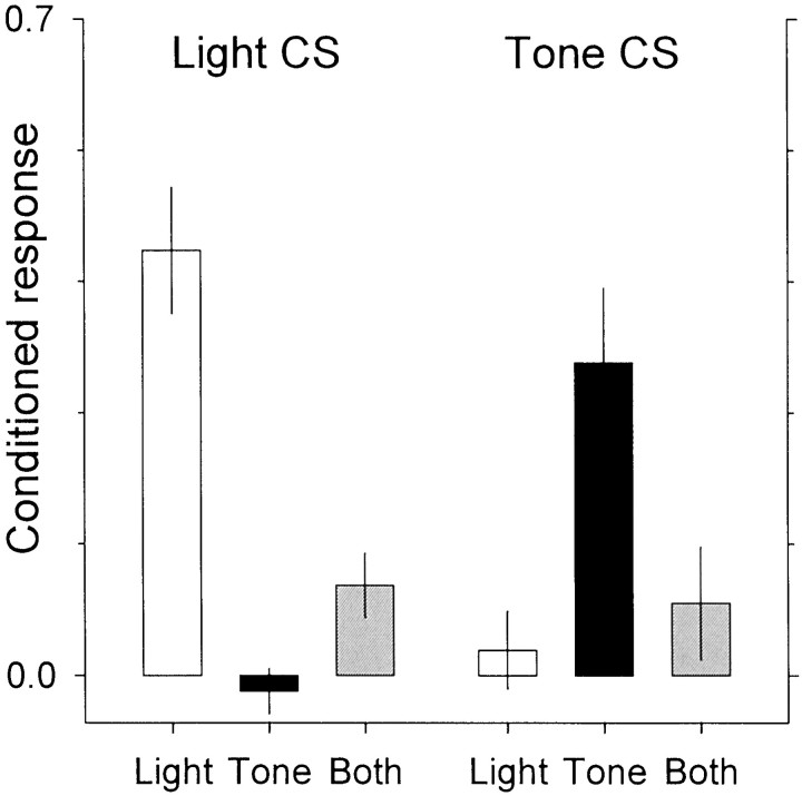
All senses, except olfaction, are routed through the thalamus to cerebral cortex. Thus, the thalamus is often referred to as the sensory gateway to cortex. Located between thalamus and cortex is a thin lamina of neurons called the thalamic reticular nucleus, which may function as an attentional gate. The phenomenon of blocking in classical conditioning provides an opportunity to test whether an attended stimulus activates the thalamic reticular nucleus more than an unattended stimulus: when a second stimulus is presented together with a previously conditioned stimulus, conditioned responding to the second stimulus is inhibited. Different groups of rats were given conditioning sessions with a single stimulus, a light or a tone, and then given conditioning sessions with compound (light and tone) stimuli. Blocking was confirmed using probe trials of single stimulus presentations. After a final test session of compound stimulus presentations, the brains were processed for the presence of Fos protein. Here we show that Fos-positive neurons were more numerous in the sector of the thalamic reticular nucleus associated with the attended conditioned stimulus than in the sector associated with the unattended stimulus. Thus, we provide evidence for an involvement of the thalamic reticular nucleus in selective attention.


Figures

Fig. 1.
Fig. 1.
Conditioned responding was assessed in probe trials in which the stimuli—light or tones—were presented alone and not followed by food reward. Responses in the 1 sec before stimulus onset were subtracted from responses during the 1 sec stimulus. Theleft panel shows responding to the light; theright panel shows responding to the tone. The rats were preconditioned, either with the light (white bars), the tone (black bars), or the compound (gray bars) stimulus. The number of hopper entries was highest when the preconditioned stimulus was presented and lowest when the blocked stimulus was presented. Intermediate values were observed for rats trained with the compound stimulus.
Fig. 2.
Fig. 2.
Mean counts of Fos-positive neurons in the dorsal 50% and ventral 50% of the thalamic reticular nucleus on sections between bregma −2.7 and −4.1 mm for each group.
Fig. 3.
Fig. 3.
Coronal sections through the thalamic reticular nucleus of the same hemisphere of one rat from each group, showing Fos-positive neurons in different sectors of the thalamic reticular nucleus according to the group. Fos staining was seen in the dorsal (visual) portion of the thalamic reticular nucleus in the light-conditioned, but not in the tone-conditioned, group. The converse pattern of activity was found in the ventral and more posterior (auditory) portion of the thalamic reticular nucleus. In the compound stimulus-conditioned rats, Fos-positive neurons were found in both regions. On the test day, all rats received the same, compound, stimulus presentations. Abbreviations from Paxinos and Watson (1997):Rt, thalamic reticular nucleus; fi, fimbria; st, stria terminalis; ic, internal capsule; eml, external medullary lamina;LDVL, laterodorsal nucleus ventrolateral division;VPM, ventral posteromedial thalamic nucleus;VPL, ventral posterolateral thalamic nucleus.
Fig. 4.
Fig. 4.
Plots showing the residual conditioned response against the residual Fos count. The visual responses (visual conditioned response and the mean Fos count in the visual portion of the thalamic reticular nucleus) were predicted from the auditory responses and vice versa. The observed responses were then subtracted from predicted responses, to obtain the residual. Thus, variability in the residuals of the visual (or auditory) responses is independent of variability in the auditory (or visual) responses.

References

    1. Coleman KA, Mitrofanis J. Organization of the visual reticular thalamic nucleus of the rat. Eur J Neurosci. 1996;8:388–404. - PubMed
    1. Cox CL, Huguenard JR, Prince DA. Nucleus reticularis neurons mediate diverse inhibitory effects in thalamus. Proc Natl Acad Sci USA. 1997;94:8854–8859. - PMC - PubMed
    1. Crick F. Function of the thalamic reticular complex: the searchlight hypothesis. Proc Natl Acad Sci USA. 1984;81:4586–4590. - PMC - PubMed
    1. Friedberg EB, Ross DT. Degeneration of rat thalamic reticular neurons following intrathalamic demoic acid injection. Neurosci Lett. 1993;151:115–119. - PubMed
    1. French CR, Sefton AJ, Mackay-Sim A. The inhibitory role of the visually responsive region of the thalamic reticular nucleus in the rat. Exp Brain Res. 1985;57:471–479. - PubMed

Publication types

Substances

LinkOut - more resources