. 2000 Dec;11(12):4105-16.
doi: 10.1091/mbc.11.12.4105.

Human orthologs of yeast vacuolar protein sorting proteins Vps26, 29, and 35: assembly into multimeric complexes

Affiliations
Free PMC article

Human orthologs of yeast vacuolar protein sorting proteins Vps26, 29, and 35: assembly into multimeric complexes

C R Haft et al. Mol Biol Cell. 2000 Dec.
Free PMC article

Abstract

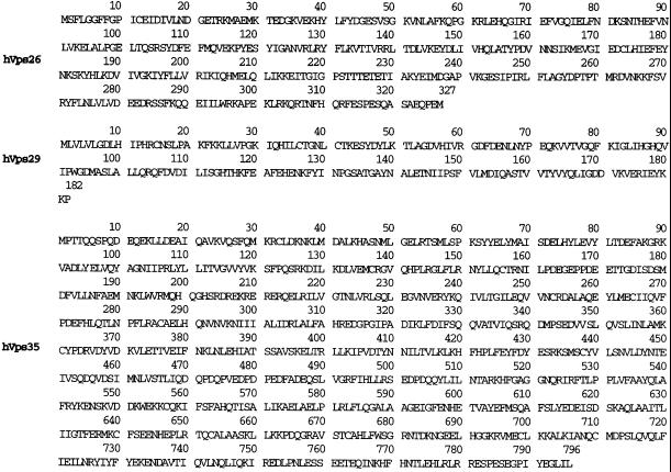
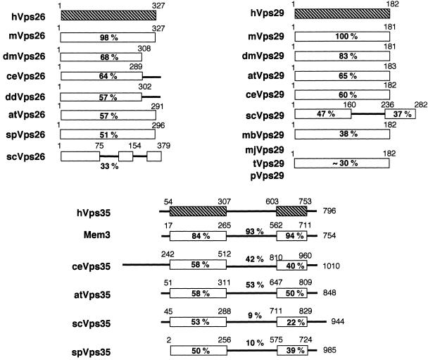
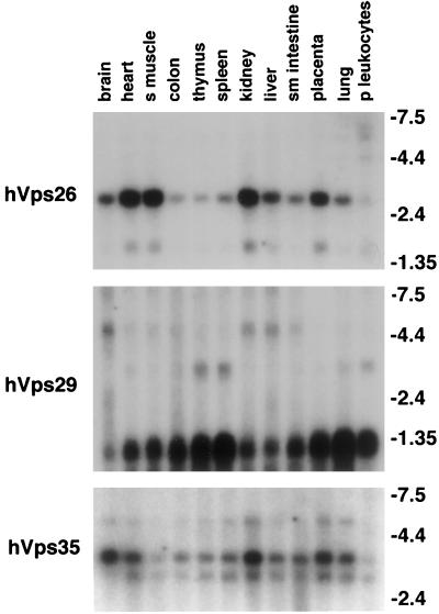
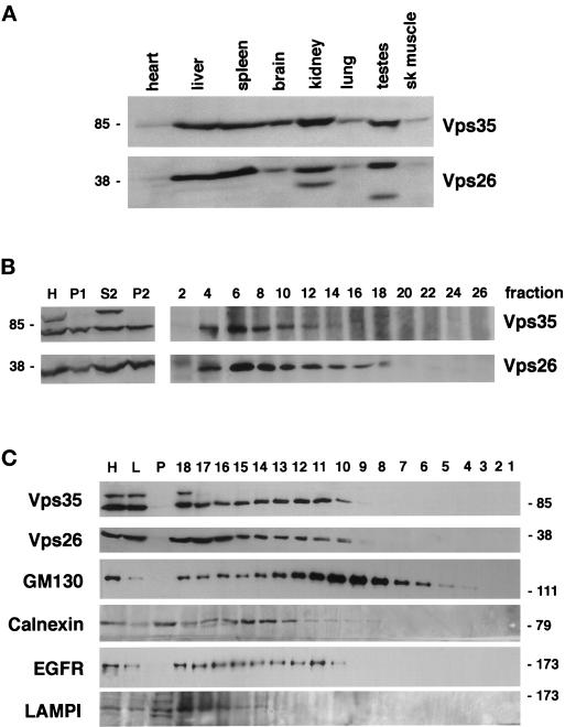
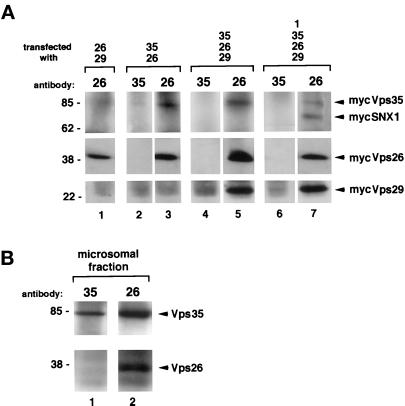
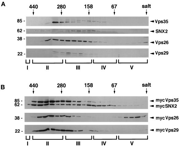
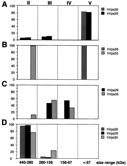
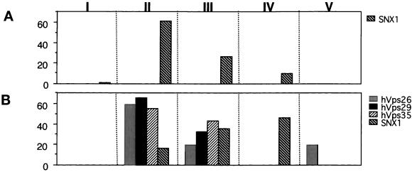
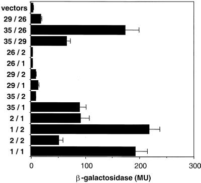
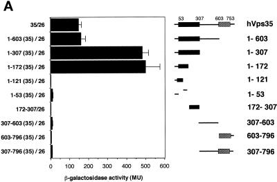
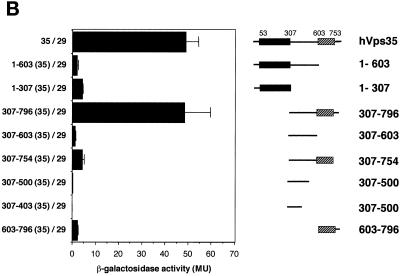
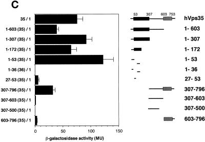
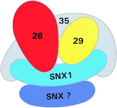
Sorting nexin (SNX) 1 and SNX2 are mammalian orthologs of Vps5p, a yeast protein that is a subunit of a large multimeric complex, termed the retromer complex, involved in retrograde transport of proteins from endosomes to the trans-Golgi network. We report the cloning and characterization of human orthologs of three additional components of the complex: Vps26p, Vps29p, and Vps35p. The close structural similarity between the yeast and human proteins suggests a similarity in function. We used both yeast two-hybrid assays and expression in mammalian cells to define the binding interactions among these proteins. The data suggest a model in which hVps35 serves as the core of a multimeric complex by binding directly to hVps26, hVps29, and SNX1. Deletional analyses of hVps35 demonstrate that amino acid residues 1-53 and 307-796 of hVps35 bind to the coiled coil-containing domain of SNX1. In contrast, hVps26 binds to amino acid residues 1-172 of hVps35, whereas hVps29 binds to amino acid residues 307-796 of hVps35. Furthermore, hVps35, hVps29, and hVps26 have been found in membrane-associated and cytosolic compartments. Gel filtration chromatography of COS7 cell cytosol showed that both recombinant and endogenous hVps35, hVps29, and hVps26 coelute as a large complex ( approximately 220-440 kDa). In the absence of hVps35, neither hVps26 nor hVps29 is found in the large complex. These data provide the first insights into the binding interactions among subunits of a putative mammalian retromer complex.


Figures

Figure 1
Figure 1
Amino acid sequences of human retromer proteins. Shown are the deduced amino acid sequences for human Vps26, Vps29, and Vps35 (accession number AF175266, AF175264, and AF175265, respectively). The predicted sizes of 36, 20, and 87 kDa for hVps26, hVps29, and hVps35, respectively, correspond very well with the size of each protein seen by Western blotting.
Figure 2
Figure 2
Human Vps26, Vps29, and Vps35 are evolutionarily well conserved. Shown are schematic illustrations of the predicted domain architecture of human (h) Vps26, Vps29, and Vps35, and related proteins from C. elegans (ce), S. cerevisiae (se), Schizosaccharomyces pombe (sp), mouse (m), A. thaliana (at), D. melanogaster (dm), Dictyostelium discoideum (dd), M. jannaschii (mj), M. thermoautotrophicum (mb), T. maritima (t), and P. abyssi (p). The accession numbers of the homologous molecules are as follows: mVps26, P40336; dmVps26, T12683, ceVps26, O01258; ddVps26, AAB03668; atVps26, T05874; spVps26, Q10243; scVps26, NP004887; mVps29, AF193794; dmVps29, AAF51410; atVps29, T07720; ceVps29, T27697; scVps29, NP011876; mjVps29, Q58040; mbVps29, O27802; tVps29, D72296; pVps29, H75143; Mem3, NP032608, ceVps35, T34314; atVps35, AAB80794; scVps35, NP012381; spVps35, T11719. The percentage of amino acid identity between each human protein and related proteins from other species as determined by CLUSTAL W alignment program (Thompson et al., 1994) is shown below the various domains of the proteins.
Figure 3
Figure 3
hVps35, hVps29, and hVps26 are ubiquitously expressed. The tissue distributions of hVps26, hVps29, and hVps35 mRNAs are shown. Filters containing poly(A)+ RNA from the indicated human tissues were hybridized with radiolabeled human Vps26, Vps29, and Vps35 probes. Single messages of ∼3 and ∼1.2 kDa were found for hVps26 and hVps29, respectively. Two mRNA species were found for hVps35. The more abundant form migrated at ∼3.4 kDa and the less abundant form at ∼3 kDa. The apparent mRNA sizes seen for hVps26, hVps29, and hVps35 are within the range expected when the 5′ and 3′ untranslated regions observed in several cDNA clones are added to the predicted coding sequence for each molecule.
Figure 4
Figure 4
hVps35, hVps29, and hVps26 are found in both cytosolic and membrane-associated pools. (A) Distribution of hVps35 and hVps26 proteins in the indicated rat tissues as determined by immunoblotting of the various tissue extracts (∼10 μg of total protein) with molecule-specific polyclonal antibodies and anti-rabbit-horseradish peroxidase. (B) Subcellular distribution of endogenous retromer proteins in various rat liver fractions and on a linear sucrose gradient through which a microsomal fraction was sedimented as described in MATERIALS AND METHODS. Aliquots of liver homogenate (H), nuclei and unbroken cells (P1), a cytosolic fraction (S2), and a microsomal pellet (P2) were analyzed by Western blotting with anti-Vps35 and anti-Vps26 polyclonal antibodies. The distribution of hVps35 and hVps26 proteins in gradient fractions collected from rat liver microsomes separated on a linear sucrose gradient (1.06–1.25 g/ml) are also shown. The indicated gradient fractions (600 μl) were immunoprecipitated with Vps35- and Vps26-specific polyclonal antibodies, and the distribution of each protein in the various fractions determined. Fractions 1–4 contained material that remained in the load, fractions 5–10 are enriched in endosomes (EEA1 positive); fractions 13–18 are enriched in Golgi membranes (GM130 positive); fractions 18–21 are enriched in plasma membrane and endoplasmic reticulum (epidermal growth factor receptor and calnexin positive, respectively); and fractions 22–30 are enriched in lysosomes (LAMPI positive) and mitochondria as determined by immunoblotting for the indicated marker proteins (our unpublished results). (C) Floatation analysis of total rat liver homogenate. An aliquot of the starting dense homogenate (H), the material remaining in the load after centrifugation (L), the material that pelleted after centrifugation, and each gradient fraction were analyzed by Western blotting with the antibodies indicated. Fraction 18 is the heaviest fraction and closest to the load, whereas fraction 1 is the lightest fraction at the top of the gradient.
Figure 5
Figure 5
Coimmunoprecipitation of hVps35, hVps29, hVps26, and SNX1. (A) COS7 cells were transiently transfected with expression vectors encoding myc-tagged Vps35, Vps26, Vps29, and SNX1 as indicated. Total cell extracts were prepared in the absence of detergent and then immunoabsorbed with the indicated polyclonal antibodies followed by protein A Sepharose. Immune complexes were collected, washed, and subjected to electrophoresis followed by immunoblotting with an anti-myc antibody. (B) Microsomal fraction prepared from rat liver was immunoabsorbed, and the samples treated as described in A, expect that the immunoblots were probed with antibodies to hVps35 and hVps26 to detect the amount of each endogenous protein that was immuno-isolated.
Figure 6
Figure 6
hVps26, hVps29, and hVps35 are found in a large complex. (A) Cell extract was prepared from nontransfected COS7 cells in the absence of detergent as described in MATERIALS AND METHODS. The extract was centrifuged at 240,000 × g for 45 min, and the resulting supernatant was chromatographed on a Sephacryl S-300 gel filtration column. The proteins in each fraction were precipitated with trichloroacetic acid, and analyzed for the presence of hVps26, hVps29, hVps35, and SNX2 by immunoblotting with antibodies made against the various proteins. The resulting autoradiograms were scanned, and the density of the bands quantified by using the NIH Image software. Arrows show the positions of marker proteins used to calibrate the column (thyroglobulin [669 kDa], ferritin [440 kDa], γ-globulin [158 kDa], bovine serum albumin [67 kDa]). (B) Cytosolic fraction prepared from COS7 cells transiently transfected with expression vectors encoding myc-tagged hVps26, hVps29, hVps35, and SNX2 was chromatographed, and the fractions treated as detailed above except that the immunoblots were probed with an anti-myc antibody to detect the overexpressed proteins. Pool I (fractions 1–13) contains molecules that elute at sizes between 103 and 440 kDa; pool II (fractions 14–18) contains molecules eluting between 440 and 280 kDa; pool III (fractions 19–24) contains molecules eluting between 280 and 158 kDa; pool IV (fractions 25–29) contains molecules eluting between 158 and 67 kDa; and pool V (fractions 30–35) contains molecules eluting at <67 kDa.
Figure 7
Figure 7
hVps29 and hVps35 form a cytosolic complex in the absence of hVps26. Cytosolic fractions were prepared from COS7 cells overexpressing cDNAs encoding myc-tagged hVps26 and hVps29 (A); hVps26 and hVps35 (B); hVps29 and hVps35 (C); and hVps26, hVps29, and hVps35 (D). Cell extracts were then prepared, chromatographed, and analyzed as described in Figure 6. Shown is the distribution of each myc-tagged molecule in the pools of various sizes. The signal detected in the various pools is expressed as a percentage of the total immunodetectable signal in all the column fractions.
Figure 8
Figure 8
SNX1 forms large oligomeric complexes in the absence the other retromer proteins. Cytosolic fractions were prepared from COS7 cells overexpressing cDNAs encoding myc-tagged SNX1 (A) and SNX1, hVps26, hVps29, and hVps35 (B). Cell extracts were then prepared, chromatographed, and analyzed as described in Figure 6. Shown is the distribution of each myc-tagged molecule in the pools of various sizes. The signal detected in the various pools is expressed as a percentage of the total immunodetectable signal in all the column fractions.
Figure 9
Figure 9
Interactions of human retromer proteins with each other and SNX1 and SNX2. Full-length SNX1, SNX2, hVps26, hVps29, and hVps35 were fused to either the LexA DNA-binding domain in the pLexA vector or the B42 activation domain in the pB42AD vector as indicated. Interactions were assessed by using a liquid β-galactosidase assay and reported in Miller units (MU). Results are presented as mean ± SE of data obtained with 12–20 independent yeast clones.
Figure 10
Figure 10
Different regions of hVps35 bind to hVps26, hVps35, and SNX1. The experiments were carried out as described in the legend to Figure 9. (A) Interactions of the pB42AD-hVps26 fusion proteins with pLexA fusion proteins containing the indicated fragments of hVps35. (B) Interactions of pB42AD-hVps29 fusion protein with pLexA fusion proteins containing the indicated fragments of hVps35. (C) Interactions of SNX1 fused to pB42AD with pLexA fusion proteins containing the indicated fragments of hVps35. Results are presented as means ± SE of data obtained with 9–20 independent yeast clones.
Figure 10
Figure 10
Different regions of hVps35 bind to hVps26, hVps35, and SNX1. The experiments were carried out as described in the legend to Figure 9. (A) Interactions of the pB42AD-hVps26 fusion proteins with pLexA fusion proteins containing the indicated fragments of hVps35. (B) Interactions of pB42AD-hVps29 fusion protein with pLexA fusion proteins containing the indicated fragments of hVps35. (C) Interactions of SNX1 fused to pB42AD with pLexA fusion proteins containing the indicated fragments of hVps35. Results are presented as means ± SE of data obtained with 9–20 independent yeast clones.
Figure 11
Figure 11
Model of binding interactions between human retromer proteins. In this model, hVps35 forms the core of a multimeric complex in which it binds directly to at least three other proteins: hVps26, hVps29, and SNX1. Furthermore, there are potential binding interactions that may stabilize the complex: hVps26 with hVps29, hVps29 with SNX1, and SNX1 with SNX1 (or another sorting nexin, designated SNX-?). In contrast, we did not detect evidence for binding of hVps26 directly to SNX 1 or SNX2. Furthermore, SNX2 does not appear to bind directly to hVps35. Although we have depicted a single molecule of SNX1 binding to both binding sites on hVps35, it is possible that each binding site on hVps35 is occupied by distinct molecules of SNX1. Our model for the mammalian retromer complex is consistent with the proposed subunit composition of the yeast retromer complex with the exception that we have not identified a human ortholog for Vps17p (see DISCUSSION).

Similar articles

Cited by

References

    1. Andersson S, Bishop RW, Russell DW. Expression cloning and regulation of steroid 5 alpha-reductase, an enzyme essential for male sexual differentiation. J Biol Chem. 1989;264:16249–16255. - PMC - PubMed
    1. Ausubel FM, Brent R, Kingston RE, Moore DD, Seidman JG, Smith JA, Struhl K. Current Protocols in Molecular Biology. Vol. 2. New York: John Wiley & Sons; 1994. pp. 13.17.11–13.17.10.
    1. Bachhawat AK, Suhan J, Jones EW. The yeast homolog of H 58, a mouse gene essential for embryogenesis, performs a role in the delivery of proteins to the vacuole. Genes Dev. 1994;8:1379–1387. - PubMed
    1. Bankaitis VA, Johnson LM, Emr SD. Isolation of yeast mutants defective in protein targeting to the vacuole. Proc Natl Acad Sci USA. 1986;83:9075–9079. - PMC - PubMed
    1. Bryant NJ, Stevens TH. Vacuole biogenesis in Saccharomyces cerevisiae: protein transport pathways to the yeast vacuole. Microbiol Mol Biol Rev. 1998;62:230–247. - PMC - PubMed

Associated data