. 2000 Dec;11(12):4117-30.
doi: 10.1091/mbc.11.12.4117.

Mutation of cyclin/cdk phosphorylation sites in HsCdc6 disrupts a late step in initiation of DNA replication in human cells

Affiliations
Free PMC article

Mutation of cyclin/cdk phosphorylation sites in HsCdc6 disrupts a late step in initiation of DNA replication in human cells

U Herbig et al. Mol Biol Cell. 2000 Dec.
Free PMC article

Abstract

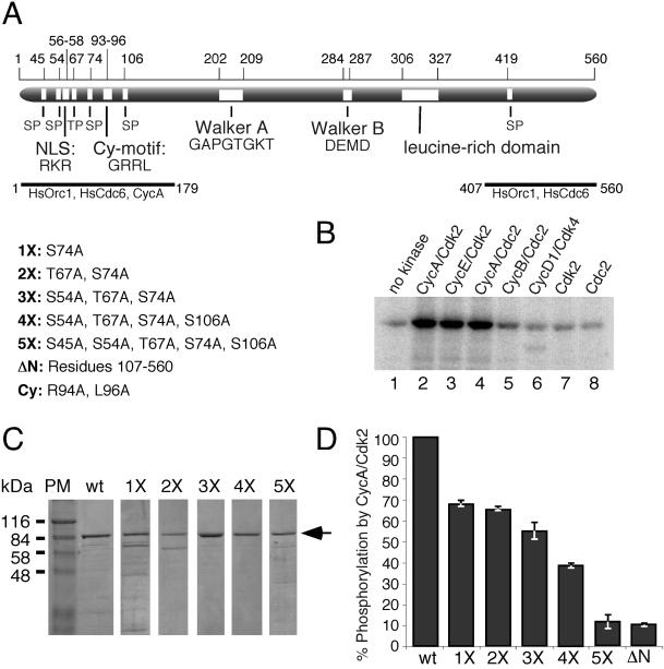
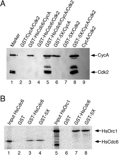
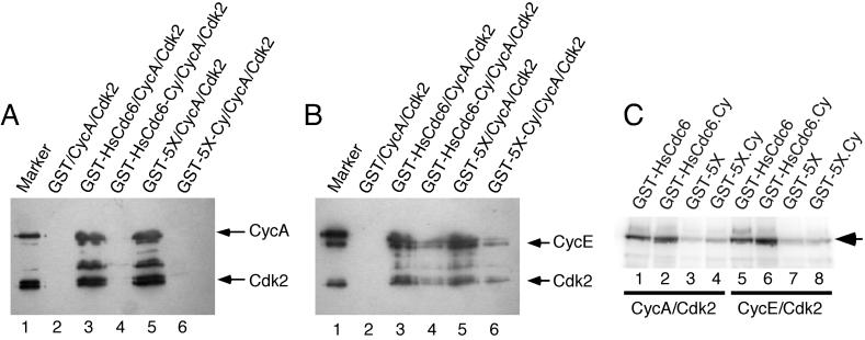
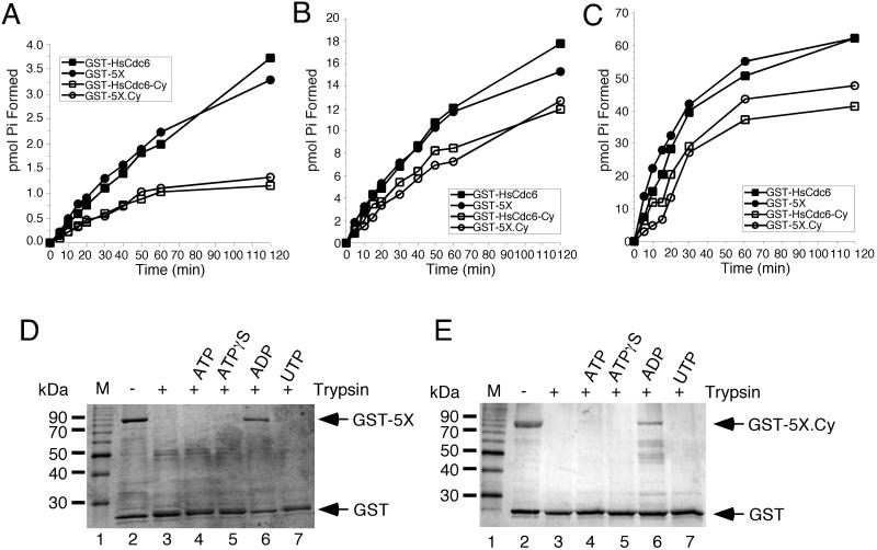
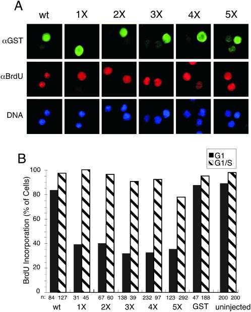
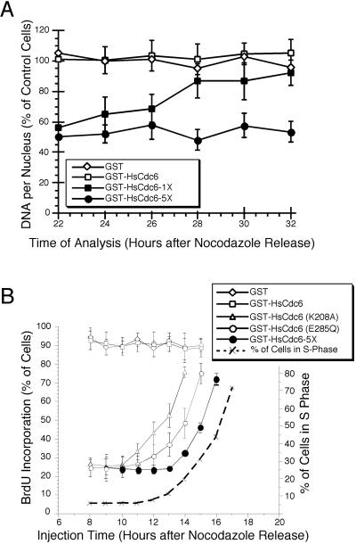
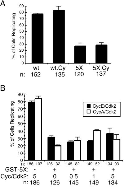
Cyclin-dependent kinases (Cdk) are essential for promoting the initiation of DNA replication, presumably by phosphorylating key regulatory proteins that are involved in triggering the G1/S transition. Human Cdc6 (HsCdc6), a protein required for initiation of DNA replication, is phosphorylated by Cdk in vitro and in vivo. Here we report that HsCdc6 with mutations at potential Cdk phosphorylation sites was poorly phosphorylated in vitro by Cdk, but retained all other biochemical activities of the wild-type protein tested. Microinjection of mutant HsCdc6 proteins into human cells blocked initiation of DNA replication or slowed S phase progression. The inhibitory effect of mutant HsCdc6 was lost at the G1/S transition, indicating that phosphorylation of HsCdc6 by Cdk is critical for a late step in initiation of DNA replication in human cells.


Figures

Figure 1
Figure 1
HsCdc6 is specifically phosphorylated at N-terminal Cdk phosphorylation sites in vitro. (A) Schematic representation of the HsCdc6 protein illustrating six potential phosphorylation sites for Cdk (SP or TP), the cyclin-binding motif (Cy-motif), the nuclear localization signal (NLS; Takei et al., 1999), the ATP binding motif (P-loop), the Mg2+ binding motif (DEAD-box), a leucine-rich domain, and peptides shown to bind to HsOrc1, HsCdc6, and cyclin A (Saha et al., 1998). The numbers above the protein indicate the position of the illustrated motifs in the amino acid sequence. Five mutant forms of HsCdc6 with amino acid substitutions at potential Cdk phosphorylation sites and combinations of sites, as indicated, were prepared and named 1X-5X; ΔN represents HsCdc6 lacking the N-terminal 106 amino acids; Cy, two alanine substitutions in the cyclin-binding motif determined previously (Petersen et al., 1999). (B) GST-HsCdc6 (3 μg) was incubated with 1 U of purified baculovirus-expressed cyclin A/Cdk2 (lane 2), cyclin E/Cdk2 (lane 3), cyclin A/Cdc2 (lane 4), cyclin B/Cdc2 (lane 6), and 1.35 U of cyclin D1/Cdk4 in the presence of [γ-32P]ATP. As a control, GST-HsCdc6 was treated similarly without kinase (lane 1) or with 0.5 μg Cdk2 alone (lane 7) or 0.5 μg Cdc2 alone (lane 8). After 10% SDS-PAGE, phosphorylated GST-HsCdc6 was visualized by PhosphorImaging. (C) Purified wild-type HsCdc6 fused to GST (wt) and GST-HsCdc6 mutants with single and multiple amino acid substitutions at potential Cdk phosphorylation sites were separated by 10% SDS-PAGE and stained with Coomassie Brilliant Blue. PM, protein marker. (D) Equal amounts of wild-type and the indicated mutant forms of GST-HsCdc6 were incubated with 6 U of purified baculovirus-expressed cyclin A/Cdk2 in the presence of [γ-32P]ATP. The proteins were resolved by 10% SDS-PAGE and visualized by Coomassie Brilliant Blue staining and PhosphorImaging (not shown). The relative amount of phosphorylation of mutant proteins compared with wild type, defined as 100%, was determined using IPLabgel software. Error bars, average error of the mean as determined from two separate experiments.
Figure 2
Figure 2
Wild-type and mutant forms of HsCdc6 bind to CycA, HsOrc1, and HsCdc6. (A) Insect cells were coinfected with recombinant baculoviruses encoding the indicated proteins. Cell extracts were prepared and incubated with glutathione agarose. After washing, the beads were boiled in sample buffer, and the eluted proteins were resolved by 10% SDS-PAGE. Cyclin A or Cdk2 that coprecipitated with the GST-fusion proteins was detected by immunoblotting using anti-CycA antibody C160 or anti-HA antibody 12CA5 to recognize the an N-terminal HA tag on Cdk2. Lane 1 contained 0.5 μg purified baculovirus-expressed CycA/Cdk2 as a marker. (B) Glutathione agarose beads containing equal amounts of GST (lanes 2 and 6), GST-HsCdc6 (lanes 3 and 7), or GST-HsCdc6-5X (GST-5X; lanes 4 and 8) were incubated with radiolabeled, in vitro–translated HsCdc6 protein (lanes 2–4) or HsOrc1 protein (lanes 6–8). After washing, the beads were boiled in sample buffer, and the eluted proteins were resolved by 10% SDS-PAGE. Bound proteins were visualized by PhosphorImaging. Lanes 1 and 5 contained formula image of the radiolabeled input protein.
Figure 3
Figure 3
HsCdc6 with mutations in the cyclin-binding motif is reduced in its affinity for cyclins, but is efficiently phosphorylated by Cdk2. (A and B) Insect cells were coinfected with recombinant baculoviruses encoding the indicated GST fusion proteins together with Cyclin A and Cdk2 (A) and Cyclin E and Cdk2 (B). Cell extracts were prepared and incubated with glutathione agarose. After washing, the beads were boiled in sample buffer, and the eluted proteins resolved by 10% SDS-PAGE. Cyclin A (A), cyclin E (B), or Cdk2 (A and B) that coprecipitated with the GST-fusion proteins was detected by immunoblotting using anti-CycA antibody C160 (A), anti-CycE antibody HE111 (B), or anti-HA antibody 12CA5 (A and B) to recognize the N-terminal HA tag on Cdk2. Lane 1 contained 0.5 μg purified baculovirus-expressed CycA/Cdk2 (A) or CycE/Cdk2 (B) as a marker. (C) Equal amounts of wild-type and the indicated mutant forms of GST-HsCdc6 were incubated with 6 U of purified baculovirus-expressed cyclin A/Cdk2 (lanes 1–4) or cyclin E/Cdk2 (lanes 5–8) in the presence of [γ-32P]ATP. The proteins were resolved by 12.5% SDS-PAGE and visualized by PhosphorImaging.
Figure 4
Figure 4
Nucleotide binding and hydrolysis properties of mutant GST-HsCdc6. (A–C) GST-HsCdc6 (0.25 pmol; ▪), GST-HsCdc6-5X (GST-5X; ●) GST-HsCdc6-Cy (□) and GST-HsCdc6-5X.Cy (GST-5X.Cy; ○) were incubated with (A) 2.5 μM, (B) 25 μM, or (C) 250 μM [γ-32P]ATP for the indicated times at 37°C. Hydrolysis products were separated by TLC, and the amount of phosphate formed was quantified by PhosphorImaging. GST-HsCdc6-5X (0.5 μg; D) or GST-HsCdc6-5X.Cy (E) bound to glutathione agarose was partially digested with trypsin in the absence of nucleotide (lane 3) or in the presence of 2 mM ATP (lane 4), ATPγS (lane 5), ADP (lane 6), or UTP (lane 7). The reaction products were analyzed by 12.5% SDS-PAGE and silver staining. No trypsin was added to the reaction shown in lane 2. M, 10-kDa marker protein ladder.
Figure 5
Figure 5
HsCdc6 with mutations at Cdk phosphorylation sites inhibits DNA replication in human cells. (A) HeLa-S3 cells were arrested for 16 h in G2/M using nocodazole. At 6–8 h after release into nocodazole-free medium, the following proteins were injected into the nucleus of the cells: GST-HsCdc6 (column 1), GST-HsCdc6-1X (1X; column 2), GST-HsCdc6-2X (2X; column 3), GST-HsCdc6-3X (3X; column 4), GST-HsCdc6-4X (4X; column 5), and GST-HsCdc6-5X (5X; column 6). The injected cells were grown for 17 h in medium containing bromodeoxyuridine (BrdU). The cells were stained with anti-GST polyclonal antibody and FITC-conjugated goat anti-rabbit secondary antibody (top row), anti-BrdU monoclonal antibody and Cy3-conjugated goat anti-mouse secondary antibody (middle row), and Hoechst 33258 fluorochrome (bottom row). Micrographs were taken at a 100× magnification with a digital camera mounted on a fluorescence microscope. (B) The following proteins at the indicated concentrations were injected into the nuclei of HeLa-S3 cells in G1 or G1/S: GST (500 ng/μl), GST-HsCdc6 (52 ng/μl), GST-HsCdc6 (1X; 23 ng/μl), GST-HsCdc6 (2X; 16.9 ng/μl), GST-HsCdc6 (3X; 53 ng/μl), GST-HsCdc6 (4X; 51 ng/μl), and GST-HsCdc6 (5X; 55.8 ng/μl). The proteins were injected either at 6–8 h after release from nocodazole (black bars) or in G1/S, immediately after release from a thymidine block (hatched bars). After injection of G1 cells, growth was continued for 17 h in medium containing BrdU. Cells injected in G1/S were grown for an additional 12 h in medium containing BrdU. Cells were fixed, stained with antibodies against GST and against BrdU, and analyzed by indirect immunofluorescence microscopy. n represents the number of cells that were successfully injected and analyzed for BrdU incorporation. Cells that incorporated BrdU as efficiently as uninjected cells were defined as BrdU-positive, whereas BrdU-negative cells were those not stained or stained weakly.
Figure 6
Figure 6
Kinetics of replication interference by mutant HsCdc6. (A) HeLa-S3 cells were arrested for 16 h in G2/M with nocodazole and released into nocodazole-free medium. At the indicated times, GST (500 ng/μl; ⋄), GST-HsCdc6 (61 ng/μl; □), GST-HsCdc6 (K208A; 20 ng/μl; ▵), GST-HsCdc6 (E285Q; 30 ng/μl; ○), and GST-HsCdc6; 5X; 55.8 ng/μl; ●) were microinjected into the nuclei of the cells. Immediately after microinjection, the medium was supplemented with BrdU, and growth was continued until 22 h after the nocodazole release. Cells were then stained with anti-GST and anti-BrdU antibodies and evaluated by indirect immunofluorescence microscopy. For each time point, the average value obtained from at least 40 injected cells and the SD of the mean are shown. The dashed line indicates the percentage of uninjected cells in S phase at the indicated times after release from a nocodazole block, as determined by flow cytometry of HeLa-S3 cells blocked and released in the same manner. (B) HeLa-S3 cells that had been arrested in G2/M with nocodazole for 16 h were released into nocodazole-free medium for 6–8 h. The following proteins at the indicated concentrations were microinjected into the nuclei of the cells: GST (500 ng/μl; ⋄), GST-HsCdc6 (61 ng/μl; □), GST-HsCdc6 (1X; 17 ng/μl; □), and GST-HsCdc6 (5X; 34 ng/μl; ○). At 12 h after the release, the medium was again supplemented with nocodazole to prevent progression through mitosis. At the indicated times, the cells were stained with anti-GST polyclonal antibody and FITC-conjugated goat anti-rabbit secondary antibody, and Hoechst 33258 fluorochrome. Nuclear DNA content of injected cells, measured by fluorescence microscopy, is expressed as a percentage of the nuclear DNA content of uninjected cells in the same field of vision, which was set to 100%. For each time point, the average value obtained from at least 10 cells is shown. Error bars, the SD of the mean.
Figure 7
Figure 7
Inhibition of DNA replication by HsCdc6-5X does not require stable protein interactions between HsCdc6-5X and G1 cyclins. (A) HeLa-S3 cells were arrested for 16 h in G2/M using nocodazole. At 6–8 h after release into nocodazole-free medium, the following proteins were injected into the nucleus of the cells: GST-HsCdc6 (wt), GST-HsCdc6-Cy (wt Cy), GST-HsCdc6-5X (5X), and GST-HsCdc6-5X.Cy (5X-Cy). Cells were fixed, stained with antibodies against GST and against BrdU, and analyzed by indirect immunofluorescence microscopy. n represents the number of cells that were successfully injected and analyzed for BrdU incorporation. Cells that visibly incorporated BrdU were defined as BrdU-positive cells, whereas BrdU-negative cells were those not stained. Error bars, the average error of the mean as determined from two separate experiments. (B) HeLa-S3 cells that had been synchronized as described in (A) were injected with GST-HsCdc6-5X (GST-5X) together with indicated amounts of cyclin/Cdk complexes, shown as molar excess over GST-HsCdc6-5X. For the first column pair, cells were injected only with cyclin/Cdk. Error bars, the average error of the mean as determined from two separate experiments.

Similar articles

Cited by

References

    1. Aparicio OM, Weinstein DM, Bell. SP. Components and dynamics of DNA replication complexes in S. cerevisiae: redistribution of MCM proteins and cdc45p during S phase. Cell. 1997;91:59–69. - PubMed
    1. Araki T, Yamamoto A, Yamada M. Accurate determination of DNA content in single cell nuclei stained with Hoechst 33258 fluorochrome at high salt concentration. Histochemistry. 1987;87:331–338. - PubMed
    1. Baum B, Nishitani H, Yanow S, Nurse P. Cdc18 transcription and proteolysis couple S phase to passage through mitosis. EMBO J. 1998;17:5689–5698. - PMC - PubMed
    1. Brown GW, Jallepalli PV, Huneycutt BJ, Kelly TJ. Interaction of the S phase regulator cdc18 with cyclin-dependent kinase in fission yeast. Proc Natl Acad Sci USA. 1997;94:6142–6147. - PMC - PubMed
    1. Calzada A, Sánchez M, Sánchez E, Bueno A. The stability of the Cdc6 protein is regulated by cyclin-dependent kinase/cyclin B complexes in Saccharomyces cerevisiae. J Biol Chem. 2000;275:9734–9741. - PubMed

Publication types

LinkOut - more resources