. 2000 Dec;11(12):4131-42.
doi: 10.1091/mbc.11.12.4131.

Complex phenotype of mice lacking occludin, a component of tight junction strands

Affiliations
Free PMC article

Complex phenotype of mice lacking occludin, a component of tight junction strands

M Saitou et al. Mol Biol Cell. 2000 Dec.
Free PMC article

Abstract

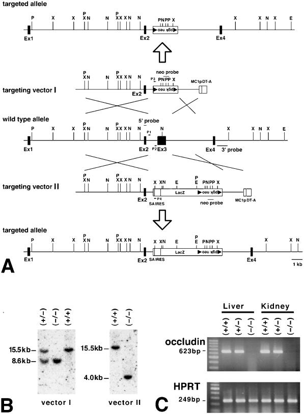
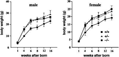
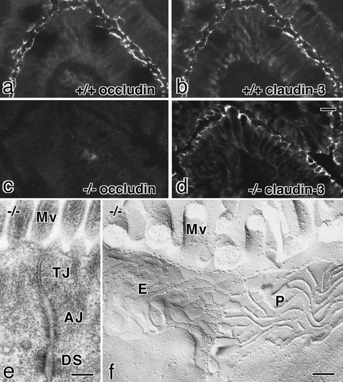
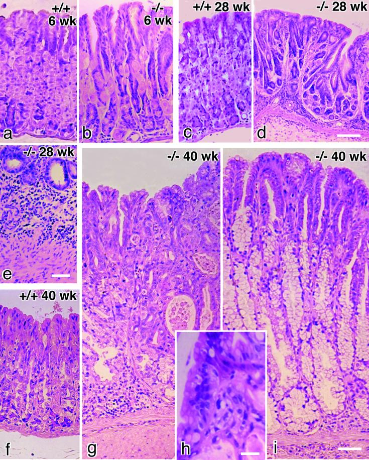
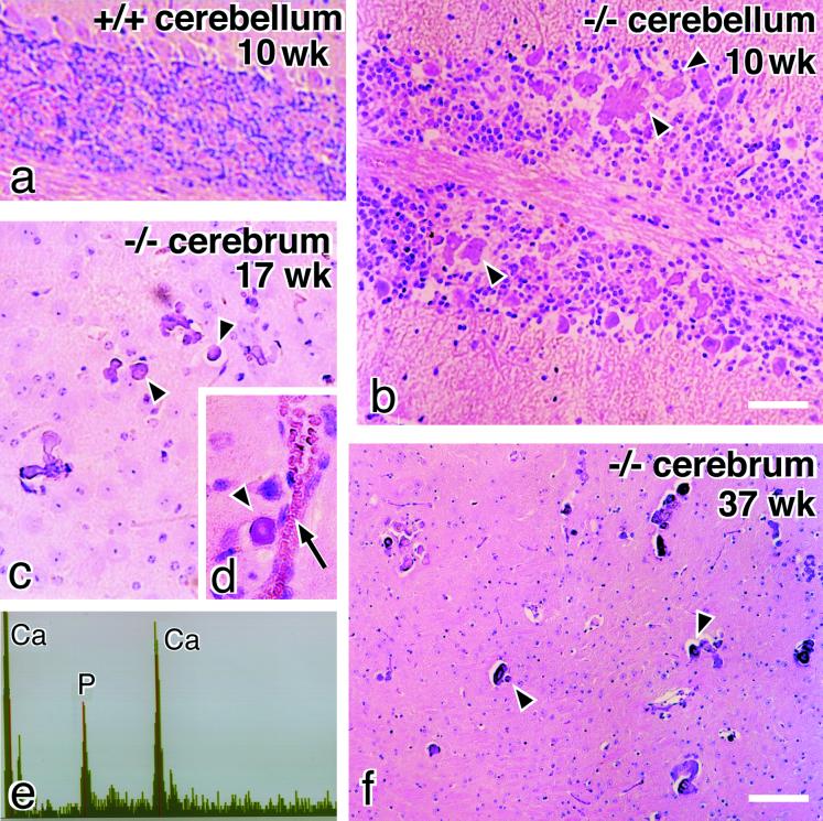
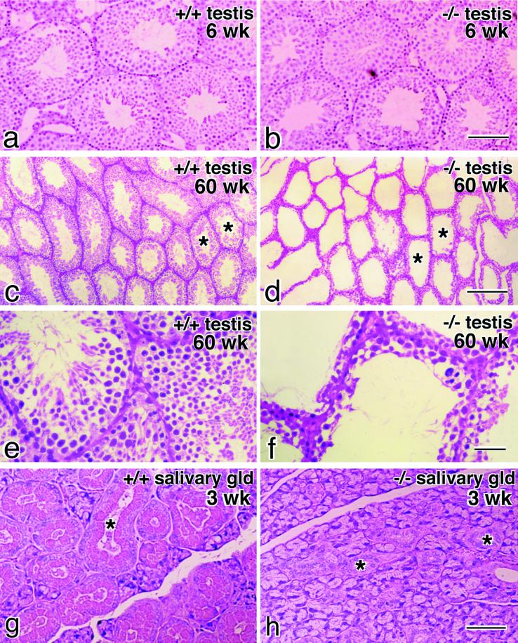
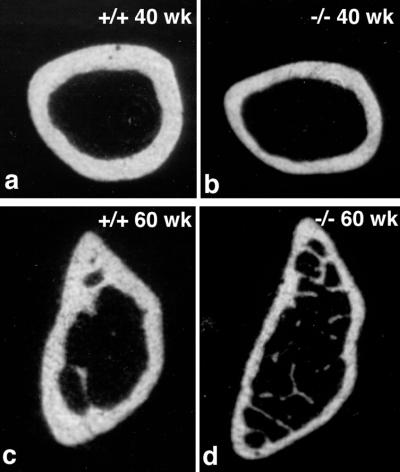
Occludin is an integral membrane protein with four transmembrane domains that is exclusively localized at tight junction (TJ) strands. Here, we describe the generation and analysis of mice carrying a null mutation in the occludin gene. Occludin -/- mice were born with no gross phenotype in the expected Mendelian ratios, but they showed significant postnatal growth retardation. Occludin -/- males produced no litters with wild-type females, whereas occludin -/- females produced litters normally when mated with wild-type males but did not suckle them. In occludin -/- mice, TJs themselves did not appear to be affected morphologically, and the barrier function of intestinal epithelium was normal as far as examined electrophysiologically. However, histological abnormalities were found in several tissues, i.e., chronic inflammation and hyperplasia of the gastric epithelium, calcification in the brain, testicular atrophy, loss of cytoplasmic granules in striated duct cells of the salivary gland, and thinning of the compact bone. These phenotypes suggested that the functions of TJs as well as occludin are more complex than previously supposed.


Figures

Figure 1
Figure 1
Generation of occludin −/− mice. (A) Restriction maps of the wild-type allele, targeting vectors I and II, and the respective targeted alleles of the mouse occludin gene. The first ATG codon was located in exon 2; exon 3 encoded the NH2-terminal half of the occludin molecule from the first transmembrane domain to the second extracellular loop. Exon 4 encoded the fourth transmembrane domain and the initial part of the COOH-terminal cytoplasmic domain, indicating the existence of one or more downstream exons. The targeting vectors I and II contained the loxP/pgk neo/loxP cassette and SA IRES/LacZ/loxP/pgk neo/loxP cassette, respectively, in their middle portion to delete exon 3 in the targeted alleles. The positions of 5′, 3′, and neo probes for Southern blotting are indicated by bars (5′ probe, 3′ probe, and neo probe), and those of primers for PCR are indicated by arrows (P1, P2, P3, P4; see MATERIALS AND METHODS). The loxP sequence is shown by closed triangles. P, PstI; X, XbaI; N, NcoI; E, EcoRV. (B) Genotype analyses by Southern blotting of XbaI-digested genomic DNA from animals that were wild-type (+/+) or either heterozygous (+/−) or homozygous (−/−) for the mutant occludin gene allele. Southern blotting with the 5′ probe yielded a 15.5-kb band, an 8.6-kb band, and a 4.0-kb band from the wild-type allele, vector I-, and vector II-targeted alleles, respectively. 3′ and neo probes were used to confirm the correct targeting. Genotyping was also performed by PCR with primers such as P1, P2, P3, and P4 (our unpublished results). (C) Loss of occludin mRNA in the liver and kidney of occludin −/− mice examined by RT-PCR. Occludin expression was abolished in occludin −/− mice, but not in wild-type or occludin +/− mice. As a control, the hypoxanthine phosphoribosyl transferase gene was equally amplified in all samples.
Figure 2
Figure 2
Postnatal growth retardation of occludin −/− mice. Occludin −/−, +/−, and +/+ mice of both sexes were born with similar body weights, but around 4 wk of age the average body weights of occludin −/− mice began to lag behind those of occludin +/− and +/+ mice (n = 7 for each). After 6 wk of age, occludin −/− mice were visibly and significantly smaller than their wild-type/heterozygous littermates.
Figure 3
Figure 3
Tight junctions in occludin −/− mice. a–d, Immunofluorescence microscopy of frozen sections of intestinal epithelial cells of occludin +/+ (a and b) and −/− (c and d) mice with anti-occludin mAb (a and c) or anticlaudin-3 polyclonal antibody (b and d). The subcellular localization of claudin-3 did not appear to be affected by the loss of occludin expression. (e and f) Ultrathin-section (e) and freeze-fracture replica (f) images of the junctional complex region of intestinal epithelial cells of occludin −/− mice. TJs and tight junction strands/grooves were indistinguishable from those in occludin +/+ mice. AJ, adherens junction; DS, desmosome; Mv, microvilli; P, P-face; E, E-face. Bars, 10 μm for a–d and 0.1 μm for e and f.
Figure 4
Figure 4
Chronic inflammation and successive hyperplasia in gastric epithelium in occludin −/− mice. Paraffin sections were stained with hematoxylin-eosin. At 6 wk of age, compared with wild-type mice (a), the gastric epithelium of occludin −/− mice was characterized by the loss of chief cells, a decrease in number of parietal cells, and the occurrence of abnormal mucoid-containing cells (b). Around 28 wk of age (c, wild-type), severe inflammatory infiltrates (e) were seen in the abnormal gastric glands with multiple branches in occludin −/− mice (d). In older occludin −/− mice around 40 wk of age, compared with wild-type mice (f) the gastric mucosa was significantly thickened (g and i). In some cases, the thickened gastric epithelium was characterized by cellular crowding, nuclear pleomorphism, increased nuclear:cytoplasmic ratio, and loss of nuclear polarity (g and h), and in other cases, the gastric glands had a pyloric gland-like appearance (i). Bars, 100 μm for a–d, f, g, and i; 40 μm for e; and 10 μm for h.
Figure 5
Figure 5
Calcification in the brain of occludin −/− mice. Paraffin sections of the cerebellum (a and b) and basal ganglia (c, d, and f) were stained with hematoxylin-eosin. Numerous concentric, often laminated mineral deposits (arrowheads) were observed in the cerebellum (b) and basal ganglia (c, d, and f) in occludin −/− mice. These deposits were often localized along small vessels (arrow in d). This kind of deposit was not found in the brain of wild-type mice (a). EDX analysis of these deposits showed calcium and phosphorus peaks similar to those of hydroxyapatite (e). Bars, 100 μm for a and b, and 200 μm for c, d, and f.
Figure 6
Figure 6
Testis and salivary gland in occludin −/− mice. Paraffin sections of the testis from 6-wk-old (a and b) or 60-wk-old mice (c–f) and the salivary gland from 3-wk-old mice (g and h) were stained with hematoxylin-eosin. In young occludin −/− mice, the testis was normally developed with normal germ cells (a and b), but in older mice the seminiferous tubules showed typical atrophy (d and f) compared with those in wild-type mice (c and e). The atrophied tubules were devoid of germ cells and retained only Sertoli cells (f). When the salivary gland of occludin −/− mice (h) was compared with that of wild-type mice (g), it was clear that striated duct cells (asterisks) in occludin −/− mice lacked cytoplasmic granules. Bars, 100 μm for a and b, 200 μm for c and d, 40 μm for e and f, and 100 μm for g and h.
Figure 7
Figure 7
X-ray computer tomography of the femur bone. Compared with wild-type mice (a and c), the compact bone was significantly thinner in occludin −/− mice (b and d).

References

    1. Anderson JM, van Itallie CM. Tight junctions and the molecular basis for regulation of paracellular permeability. Am J Physiol. 1995;269:G467–G475. - PubMed
    1. Ando-Akatsuka Y, Saitou M, Hirase T, Kishi M, Sakakibara A, Itoh M, Yonemura S, Furuse M, Tsukita Sh. Interspecies diversity of the occludin sequence: cDNA cloning of human, mouse, dog, and rat-kangaroo homologues. J Cell Biol. 1996;133:43–47. - PMC - PubMed
    1. Balda MS, Whitney JA, Flores C, González S, Cereijido M, Matter K. Functional dissociation of paracellular permeability and transepithelial electrical resistance and disruption of the apical-basolateral intramembrane diffusion barrier by expression of a mutant tight junction membrane protein. J Cell Biol. 1996;134:1031–1049. - PMC - PubMed
    1. Chen Y-H, Merzdorf C, Paul DL, Goodenough DA. COOH terminus of occludin is required for tight junction barrier function in early Xenopus embryos. J Cell Biol. 1997;138:891–899. - PMC - PubMed
    1. Chomczynski P, Sacchi N. Single-step method of RNA isolation by acid guanidinium thiocyanate-phenol-chloroform extraction. Anal Biochem. 1987;162:156–159. - PubMed

Publication types

MeSH terms