. 2000 Dec;11(12):4159-71.
doi: 10.1091/mbc.11.12.4159.

Self-association of coilin reveals a common theme in nuclear body localization

Affiliations
Free PMC article

Self-association of coilin reveals a common theme in nuclear body localization

M D Hebert et al. Mol Biol Cell. 2000 Dec.
Free PMC article

Abstract

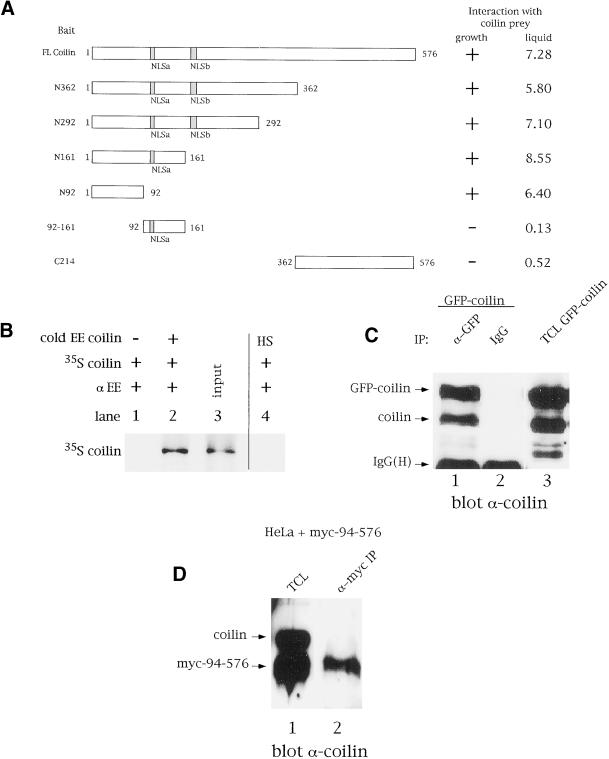
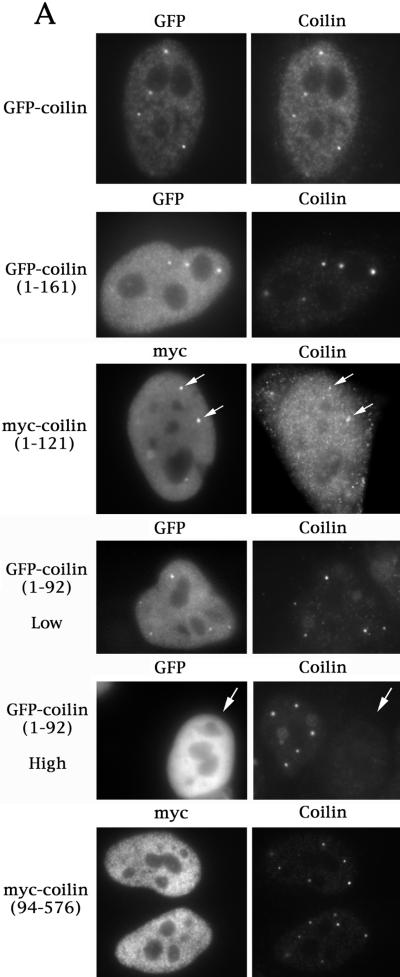
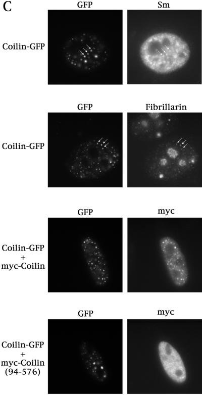
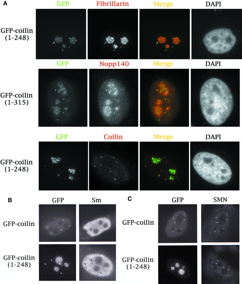
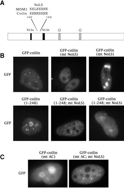
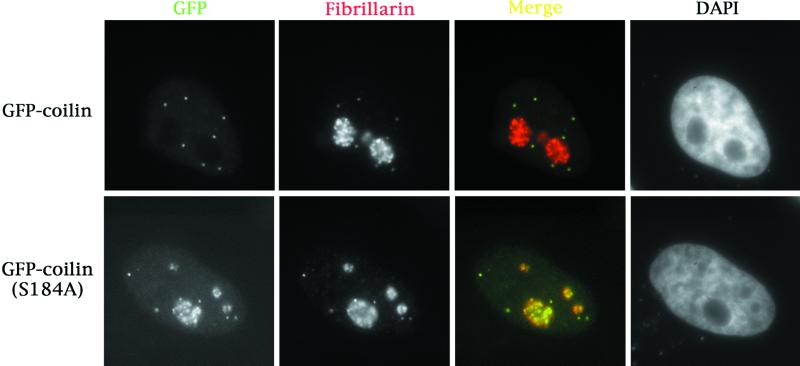
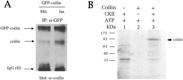
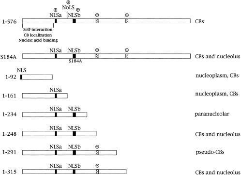
We have found that coilin, the marker protein for Cajal bodies (coiled bodies, CBs), is a self-interacting protein, and we have mapped the domain responsible for this activity to the amino-terminus. Together with a nuclear localization signal, the self-interaction domain is necessary and sufficient for localization to CBs. Overexpression of various wild-type and mutant coilin constructs in HeLa cells results in disruption of both CBs and survival motor neurons (SMN) gems. Additionally, we have identified a cryptic nucleolar localization signal (NoLS), within the coilin protein, which may be exposed in specific coilin phospho-isoforms. The implications of these findings are discussed in light of the fact that other proteins known to localize within nuclear bodies (e. g., PML, SMN and Sam68) can also self-associate. Thus protein self-interaction appears to be a general feature of nuclear body marker proteins.


Figures

Figure 1
Figure 1
The N-terminus of coilin mediates self-interaction. Coilin self-interacts in the yeast two-hybrid system (A), as well as by coimmunoprecipitation in vitro (B) and from cell extracts (C). In panel A, various coilin baits, indicated on the left, were cotransformed into the yeast strain PJ69–2A with coilin prey. Interaction was defined as the ability of PJ69–2A containing both bait and prey to grow on selective medium within 3 days. The (+) sign denotes robust growth after 3 days incubation, whereas the ({minus}) sign represents no growth. The lack of coilin prey interaction with aa 92–161 or the C-terminal 214 aa of coilin verified the specificity of the interaction. The interactions were quantified by performing liquid β-galactosidase assays, as described in MATERIALS AND METHODS. Values are reported in Miller units and varied by no more than 20%. (B) Coilin and EE-coilin were in vitro translated with or without [35S]methionine, respectively. Cold EE-coilin (5 μl) was mixed with 5 μl 35S-labeled coilin and immunoprecipitated with anti-EE antibodies (lane 2). Lane 3 contains 1/15th the amount of 35S-labeled coilin used in the reactions. No 35S-labeled coilin was recovered when immunoprecipitated in the absence of EE-coilin (lane 1) or in the presence of an EE-tagged heterologous sequence (HS, lane 4). (C) Cell extracts from GFP-coilin transfected HeLa cells were subjected to immunoprecipitation with anti-GFP antibodies, followed by SDS-PAGE and western blotting with antibodies against coilin. Normal rabbit serum was used as a negative control for immunoprecipitation. Lane 3 contains 10 μl of total cell lysate (TCL) from GFP-coilin transfected cells. IgG(H) is the immunoglobulin heavy chain. (D) Cell extracts from myc-coilin(94–576) transfected HeLa cells were immunoprecipitated with antimyc antibodies, followed by SDS-PAGE and western blotting with antibodies against coilin. No endogenous coilin is observed in lane 2, corresponding to the IP using antimyc antibodies. Lane 1 contains 10 μl of total cell lysate (TCL) from myc-coilin(94–576) transfected cells. Normal mouse serum was used as a negative control for immunoprecipitation (our unpublished results).
Figure 2
Figure 2
(A) The self-interaction domain of coilin is necessary and sufficient for CB localization. HeLa cells transfected with the indicated constructs were assayed for coilin localization by immunofluorescence using anticoilin antibody R508. The myc-coilin(1–121) and myc-coilin(94–576) constructs (Bohmann et al., 1995) were localized using antimyc antibodies. GFP-myc-NLS-coilin(1–92), labeled GFP-coilin(1–92), is shown at both high and low expression levels. The highly expressing GFP-myc-NLS-coilin(1–92) cell is indicated by an arrow. GFP-coilin(1–161) and myc-coilin(1–121) are shown at low expression levels. Facing page: (B) Overexpression of GFP-coilin disrupts CBs and results in the accumulation of GFP-coilin in thenucleolus. GFP-coilin was transfected into HeLa cells and immunofluorescence with coilin, SMN, and PML antibodies was conducted. (C) Fusion of GFP to the C-terminus of coilin apparently increases the number of CBs. Coilin was cloned into pEGFP-N3 (Clontech) and transfected into HeLa cells alone or in the presence of myc-coilin or myc-coilin (94–576). Immunofluorescence with antibodies to Sm (for snRNP localization), fibrillarin (for snoRNPs) or myc was conducted. Coilin-GFP localizes to numerous small foci which colocalize with Sm, fibrillarin, and ectopically expressed coilin, some of which are indicated by arrows. No colocalization was observed for coilin-GFP with myc-coilin(94–576).
Figure 2
Figure 2
(A) The self-interaction domain of coilin is necessary and sufficient for CB localization. HeLa cells transfected with the indicated constructs were assayed for coilin localization by immunofluorescence using anticoilin antibody R508. The myc-coilin(1–121) and myc-coilin(94–576) constructs (Bohmann et al., 1995) were localized using antimyc antibodies. GFP-myc-NLS-coilin(1–92), labeled GFP-coilin(1–92), is shown at both high and low expression levels. The highly expressing GFP-myc-NLS-coilin(1–92) cell is indicated by an arrow. GFP-coilin(1–161) and myc-coilin(1–121) are shown at low expression levels. Facing page: (B) Overexpression of GFP-coilin disrupts CBs and results in the accumulation of GFP-coilin in thenucleolus. GFP-coilin was transfected into HeLa cells and immunofluorescence with coilin, SMN, and PML antibodies was conducted. (C) Fusion of GFP to the C-terminus of coilin apparently increases the number of CBs. Coilin was cloned into pEGFP-N3 (Clontech) and transfected into HeLa cells alone or in the presence of myc-coilin or myc-coilin (94–576). Immunofluorescence with antibodies to Sm (for snRNP localization), fibrillarin (for snoRNPs) or myc was conducted. Coilin-GFP localizes to numerous small foci which colocalize with Sm, fibrillarin, and ectopically expressed coilin, some of which are indicated by arrows. No colocalization was observed for coilin-GFP with myc-coilin(94–576).
Figure 3
Figure 3
Two coilin truncation mutants localize to the nucleolus and CBs, but do not mis-localize endogenous coilin, snRNPs, or SMN to the nucleolus. (A) GFP-coilin(1–248) and GFP-coilin(1–315) mutants, upon transfection into HeLa, were found to have identical nucleolar and CB expression profiles. Antibodies to fibrillarin and Nopp140, which normally localize in nucleoli and CBs, reveal complete colocalization with these proteins. Endogenous coilin localization was determined in GFP-coilin(1–248) transfected cells using anticoilin antibodies that recognize epitopes downstream of the truncation. Note that endogenous coilin is not mislocalized to the nucleolus in GFP-coilin(1–248)transfected cells. (B and C) GFP-coilin(1–248) transfected cells were assessed for the localization of Sm proteins (B) and SMN (C). No nucleolar accumulations of snRNPs or SMN were observed. As expected, accumulations of snRNPs could be detected in the CBs (arrows). Likewise, SMN is present in the CB foci of GFP-coilin(1–248) transfected cells (our unpublished results). However, a small fraction of GFP-coilin(1–248) cells did not display CBs, yet SMN foci, or gems, are present in these cells (C).
Figure 4
Figure 4
Identification and mutation of a nucleolar localization signal (NoLS) in coilin. (A) Coilin contains a sequence, located between the two putative NLSs, that is nearly identical to the NoLS of MDM2 (Lohrum et al., 2000). Stippled boxes indicate potentially-acidic serine patches (AC) spanning amino acids 242–259 and 312–325. Facing page: (B) The putative coilin NoLS was mutated from KKNKRKNK to IINNIINI (mt NoLS) in both the GFP-coilin and GFP-coilin(1–248) backgrounds. Note that, while the truncation alone localized to nucleoli and CBs, mutation of the NoLS depleted coilin from nucleoli. (C) Similarly, block substitution of the two acidic patches (S245–252A and S314–320A, termed GFP-coilin(mt AC)) also resulted in nucleolar accumulation. Additional localization in CBs was occasionally observed (our unpublished results). However, mutant proteins bearing substitutions in both the NoLS and the acidic patches (GFP-coilin(mt AC; mt NoLS)) did not accumulate in nucleoli, resulting in a distribution pattern indistinguishable from that of wild-type coilin.
Figure 5
Figure 5
Mutation of the CDK2/cyclin E phosphorylation site in coilin at S184 to alanine results in nucleolar and CB localization. GFP-coilin(S184A) was transfected into HeLa cells. Immunofluorescence with antibodies to fibrillarin showed complete colocalization with GFP-coilin(S184A). Approximately 30% of transfected cells show this phenotype; the rest of the cells appear normal.
Figure 6
Figure 6
(A) Hyperphosphorylation of coilin reduces coilin self-interaction. GFP-coilin was transfected into HeLa and cells were treated with nocodazole to enrich for mitotic cells (Mit.) or left untreated for interphase cells (Int.). Extracts were subjected to immunoprecipitation with anti-GFP antibodies, followed by SDS-PAGE and western blotting with antibodies against coilin. Equivalent amounts of protein were used for each reaction. (B) Coilin was phosphorylated by casein kinase 2 in vitro. Reactions were conducted as described in the MATERIALS AND METHODS section. Phosphorylation of coilin was dependent on the presence of casein kinase 2 (CKII). Radiolabeled bands present in lane 1 and below the coilin radioactive band in lane 3 are most likely CKII autophosphorylation products.
Figure 7
Figure 7
Coilin mutational summary. The localization of full-length coilin (1–576) and coilin mutants generated in this study are described. The putative coilin nucleolar localization signal (NoLS), nuclear localization signals (NLS, dark boxes), self-interaction, CB localization, and nucleic acid binding domains are indicated. The NLS in the coilin(1–92) construct was added exogneously, along with GFP- and myc-tags. Positive signs (+) denote basic regions of coilin, whereas negative signs (−) indicate potentially-acidic serine patches from amino acids 242–259 and 312–325 (stippled boxes). Two additional coilin mutants, myc-coilin(1–234) and myc-coilin(1–291) were generated by Bohmann et al. (1995).

References

    1. Andrade LEC, Chan EKL, Raska I, Peebles CL, Roos G, Tan EM. Human autoantibody to a novel protein of the nuclear coiled body: Immunological characterization and cDNA cloning of p80 coilin. J Exp Med. 1991;173:1407–1419. - PMC - PubMed
    1. Andrade LEC, Tan EM, Chan EKL. Immunocytochemical analysis of the coiled body in the cell cycle and during cell proliferation. Proc Natl Acad Sci USA. 1993;90:1947–1951. - PMC - PubMed
    1. Bellini M, Gall JG. Coilin can form a complex with the U7 small nuclear ribonucleoprotein. Mol Biol Cell. 1998;9:2987–3001. - PMC - PubMed
    1. Bohmann K, Ferreira J, Lamond A. Mutational analysis of p80 coilin indicates a functional interaction between coiled bodies and the nucleolus. J Cell Biol. 1995;131:817–831. - PMC - PubMed
    1. Cajal SRy. Un sencillo metodo de coloracion selectiva del reticulo protoplasmico y sus efectos en los diversos organos nerviosos de vertebrados y invertebrados. Trab Lab Invest Biol (Madrid) 1903;2:129–221.

Publication types

LinkOut - more resources