. 2000 Dec;11(12):4277-94.
doi: 10.1091/mbc.11.12.4277.

The sodium/proton exchanger Nhx1p is required for endosomal protein trafficking in the yeast Saccharomyces cerevisiae

Affiliations
Free PMC article

The sodium/proton exchanger Nhx1p is required for endosomal protein trafficking in the yeast Saccharomyces cerevisiae

K Bowers et al. Mol Biol Cell. 2000 Dec.
Free PMC article

Abstract

We show that the vacuolar protein sorting gene VPS44 is identical to NHX1, a gene that encodes a sodium/proton exchanger. The Saccharomyces cerevisiae protein Nhx1p shows high homology to mammalian sodium/proton exchangers of the NHE family. Nhx1p is thought to transport sodium ions into the prevacuole compartment in exchange for protons. Pulse-chase experiments show that approximately 35% of the newly synthesized soluble vacuolar protein carboxypeptidase Y is missorted in nhx1 delta cells, and is secreted from the cell. nhx1 delta cells accumulate late Golgi, prevacuole, and lysosome markers in an aberrant structure next to the vacuole, and late Golgi proteins are proteolytically cleaved more rapidly than in wild-type cells. Our results show that efficient transport out of the prevacuolar compartment requires Nhx1p, and that nhx1 delta cells exhibit phenotypes characteristic of the "class E" group of vps mutants. In addition, we show that Nhx1p is required for protein trafficking even in the absence of the vacuolar ATPase. Our analysis of Nhx1p provides the first evidence that a sodium/proton exchange protein is important for correct protein sorting, and that intraorganellar ion balance may be important for endosomal function in yeast.


Figures

Figure 1
Figure 1
Deletion of the NHX1 gene. The NHX1 ORF is located on S. cerevisiae chromosome IV. YDR455C, a small yeast ORF of unknown function, overlaps NHX1 on the other DNA strand. Replacement of NHX1 with Kanr by homologous recombination also disrupts YDR455C.
Figure 2
Figure 2
Secretion of CPY in wild-type (WT), nhx1Δ::Kanr, and vps44 mutant strains, and complementation by NHX1 and NHX1-HA. Cells were labeled with [35S]methionine and cysteine for 10 min, and then chased for 0 or 40 min at 30°C. CPY was immunoprecipitated from intracellular (I) and extracellular (E) fractions for each time point. The positions of ER and Golgi precursor forms of CPY (p1 and p2, respectively), and mature vacuolar CPY (m) are indicated. nhx1Δ cells were transformed with NHX1 and YDR455C on a CEN plasmid (NHX1+YDR455C, pKEB37), or NHX1 alone with an HA epitope tag (NHX1-HA, pKEB38). vps44 cells were transformed with a CEN plasmid containing NHX1 alone with an HA-epitope tag (NHX1-HA, pKEB53). WT and nhx1Δ strains have a wild-type PEP4 gene, encoding proteinase A (required for proteolytic processing). vps44 (vpl27-1) has the pep4-3 mutation, and was transformed with PEP4 on a CEN plasmid (pTS18) for this experiment. The percentage of CPY secreted into the extracellular medium after a 40-min chase was calculated by phosphoimager analysis, and the numbers shown are averages over several separate experiments.
Figure 3
Figure 3
nhx1Δ cells have a similar morphological phenotype to vps27Δ cells. Immunofluorescence was performed as described in MATERIALS AND METHODS in wild-type (WT; SF838–9Dα), nhx1Δ (KEBY15), and vps27Δ cells (KEBY12), by using monoclonal antibodies to ALP or Pep12p, and polyclonal antibodies to Vps10p, Vph1p, or Vma2p. Staining for Vps10p, Pep12p, and ALP was enhanced by using secondary anti-mouse or anti-rabbit biotin-conjugated antibodies followed by a tertiary streptavidin-FITC conjugated antibody. Vph1p and Vma2p were detected by using a secondary Alexa-conjugated anti-rabbit antibody.
Figure 4
Figure 4
Colocalization of Pep12p, Vph1p, and Vps10p in nhx1Δ cells. Immunofluorescence was performed as described in MATERIALS AND METHODS, except cells were permeablized with 5% SDS for 5 min. The strain used was KEBY15 (nhx1Δ). Pep12p was visualized by using a monoclonal antibody against Pep12p followed by anti-mouse biotin and streptavidin-FITC. The same cells were also stained for Vps10p, or Vph1p by using polyclonal antibodies and a secondary Alexa-anti-rabbit antibody. Confocal micrographs were taken simultaneously of the red and green fluorescence channels and overlapped to produce the merged image.
Figure 5
Figure 5
FM4-64 and Ste3p-GFP accumulate in the class E compartment of nhx1Δ cells. Wild-type (WT; SF838-9Dα), nhx1Δ (KEBY15), and vps27Δ cells (SGY73) were transformed with pJLU34. Cells were labeled in 40 μM FM4-64 for 15 min at 30°C and then chased in fresh medium for 30 min at 30°C. FM4-64 and Ste3p-GFP were photographed under the red and green fluorescence channels, respectively, and a merged image of these two channels is also shown. Differential interference contrast (DIC) images of the same cells were collected to visualize the vacuoles.
Figure 6
Figure 6
Pulse-chase immunoprecipitations of Vps10p, A-ALP, and ALP. Wild-type (WT; RPY10), nhx1Δ (KEBY13), and vps27Δ cells (AACY5) were pulsed for 10 min at 30°C with [35S]cysteine and methionine, and chased for 0, 30, 60, or 120 min with excess unlabeled cysteine and methionine. Vps10p immunoprecipitates were separated on a 7% SDS polyacrylamide gel. The positions of mature Vps10p (m) and the smaller proteolytically cleaved form (*) are shown. A-ALP immunoprecipitations were carried out under the same conditions as used for Vps10p. The following strains were used, all of which had a deletion of the PHO8 gene, which encodes ALP, and were transformed with pSN55 (A-ALP on a CEN-based plasmid): BLY1 (WT), KEBY14 (nhx1Δ), and KEBY37 (vps27Δ). The positions of the precursor form of A-ALP (p) and the proteolytically cleaved form (m) are indicated. ALP immunoprecipitations were carried out as for Vps10p, but by using 0-, 5-, and 15-min chase times. The positions of precursor ALP (p) and mature ALP (m) are shown.
Figure 7
Figure 7
Analysis of the trafficking defect of vmaΔ and gef1Δ cells. (A) Proposed ion transporters of the PVC. The V-ATPase actively pumps protons into the PVC, while Nhx1p passively exchanges sodium ions for protons driven by ion gradients across the membrane. Gef1p is a putative voltage-gated ion channel thought to transport chloride ions into the PVC. (B) CPY processing and secretion. Wild-type (RPY10), nhx1Δ (KEBY13), vma2Δ (KEBY27), and gef1Δ cells (KEBY32) were labeled with [35S]methionine and cysteine for 10 min and then chased for 0 or 40 min at 30°C. CPY was immunoprecipitated as described in Figure 1, and in the MATERIALS AND METHODS. The amount of CPY secreted after a 40-min chase was quantified by using phosphoimager analysis, and is shown underneath the gel. The % CPY secreted for each strain is the average value obtained over several independent experiments (7 for WT and nhx1Δ, 3 for vma2Δ, and 2 for gef1Δ). (C) Proteolytic cleavage of Vps10p. Wild-type (RPY10), nhx1Δ (KEBY13), vma2Δ (KEBY27), vma3Δ (KEBY29 with pTS18), gef1Δ cells (KEBY32), and nhx1Δ vma2Δ cells (KEBY35) were metabolically labeled as described above and chased for 0, 30, 60, or 120 min. Vps10p immunoprecipitates were separated on a SDS polyacrylamide gel, which was exposed to a phosphoimager screen and the band intensities quantified. The amount of the lower, cleaved form of Vps10p (*) was calculated as a percentage of total Vps10p at each time point. Each point on the graph represents the mean of several independent experiments (n = number of experiments), and the error bars represent SDs from the mean.
Figure 7
Figure 7
Analysis of the trafficking defect of vmaΔ and gef1Δ cells. (A) Proposed ion transporters of the PVC. The V-ATPase actively pumps protons into the PVC, while Nhx1p passively exchanges sodium ions for protons driven by ion gradients across the membrane. Gef1p is a putative voltage-gated ion channel thought to transport chloride ions into the PVC. (B) CPY processing and secretion. Wild-type (RPY10), nhx1Δ (KEBY13), vma2Δ (KEBY27), and gef1Δ cells (KEBY32) were labeled with [35S]methionine and cysteine for 10 min and then chased for 0 or 40 min at 30°C. CPY was immunoprecipitated as described in Figure 1, and in the MATERIALS AND METHODS. The amount of CPY secreted after a 40-min chase was quantified by using phosphoimager analysis, and is shown underneath the gel. The % CPY secreted for each strain is the average value obtained over several independent experiments (7 for WT and nhx1Δ, 3 for vma2Δ, and 2 for gef1Δ). (C) Proteolytic cleavage of Vps10p. Wild-type (RPY10), nhx1Δ (KEBY13), vma2Δ (KEBY27), vma3Δ (KEBY29 with pTS18), gef1Δ cells (KEBY32), and nhx1Δ vma2Δ cells (KEBY35) were metabolically labeled as described above and chased for 0, 30, 60, or 120 min. Vps10p immunoprecipitates were separated on a SDS polyacrylamide gel, which was exposed to a phosphoimager screen and the band intensities quantified. The amount of the lower, cleaved form of Vps10p (*) was calculated as a percentage of total Vps10p at each time point. Each point on the graph represents the mean of several independent experiments (n = number of experiments), and the error bars represent SDs from the mean.
Figure 8
Figure 8
Unlike nhx1Δ or nhx1Δ vma2Δ cells, vma2Δ cells do not show a class E Vps morphological phenotype. Immunofluorescence was performed as described in Figure 4, with wild-type (WT: SF8389Dα), nhx1Δ (KEBY15), vma2Δ (KEBY26), and nhx1Δ vma2Δ (KEBY34) cells and anti-Pep12p and anti-Vph1p antibodies. Images were captured by using a fluorescence microscope fitted with a digital camera.
Figure 9
Figure 9
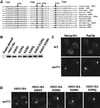
Mutation of conserved acidic residues in Nhx1p. (A) Partial sequence alignment of NHE protein sequences. NHE proteins with homology to Nhx1p have been found in many organisms. Those shown here are baker's yeast (S. cerevisiae) Nhx1p; fission yeast (S. pombe) NHE; thale cress (A. thaliana) NHX1; rice (Oryza sativa) NHX1; fruit fly (Drosophila melanogaster) NHE1; human (Homo sapiens) NHE proteins 1, 2, 3, 5, and 6; and rat (Rattus norvegicus) NHE4. The alignment was generated by using complete amino acid sequences, though only predicted transmembrane domains 5, 6, and 9 are shown. Acidic residues conserved throughout the NHE family (D201, E225, and D230 in Nhx1p) are in bold type. (B) NHX1-HA point mutant constructs make full-length proteins, expressed to wild-type levels. Whole-cell lysates were prepared from nhx1Δ cells (KEBY10) transformed with pRS316 (empty plasmid), or pRS316 with wild-type or mutant forms of NHX1-HA (pKEB38, 44, 45, 46, 47, pFP1, or pFP2). Fifteen micrograms of total protein was loaded per lane. Although E225Q appears to express slightly less Nhx1p-HA than the other strains, this was not significant over several experiments. In addition, this strain has the same point mutation as the D201N E225Q mutant, which shows wild-type expression levels in this experiment. (C) Localization of Nhx1p-HA. Rabbit polyclonal antibodies against the HA epitope were used to immunolocalize Nhx1p-HA in wild-type cells (WT; KEBY11 with pKEB38), or vps27Δ cells (vps27Δ; KEBY12 with pKEB38). Cells were costained by using a monoclonal antibody to Pep12p. (D) Localization of Nhx1p-HA point mutants. vps27Δ cells were transformed with pRS316 containing wild-type or mutant forms of NHX1-HA (pKEB38, 44, 45, 46, or 47). Nhx1p-HA proteins were immunolocalized with rabbit polyclonal antibodies against the HA epitope. The images in C and D are confocal micrographs.
Figure 10
Figure 10
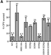
Phenotype of nhx1Δ cells expressing Nhx1p-HA point mutants. The strains used in these experiments were wild-type (WT; SEY6210 with empty plasmid), nhx1Δ (KEBY10 with empty plasmid), nhx1Δ with NHX1-HA on a CEN plasmid (NHX1-HA; KEBY10 with pKEB38), and nhx1Δ with NHX1-HA point mutants on CEN plasmids (D201N, E225Q, D230N, E355Q, D201N E225Q, and D201N D230N; KEBY10 with pKEB44, pKEB45, pKEB46, pKEB47, pFP1, and pFP2, respectively). (A) CPY secretion. Cells were labeled in [35S]cysteine and methionine for 10 min and incubated in excess nonradioactive cysteine and methionine for 40 min. CPY was immunoprecipitated as described in Figure 2 and the MATERIALS AND METHODS. The gel was exposed to a phosphoimager screen, and the amount of CPY secreted into the extracellular medium was quantified as the percentage of total CPY in the sample. Each bar on the graph represents the mean percentage of CPY secreted over five separate experiments for WT, nhx1Δ, and NHX1-HA; three separate experiments for D201N, E225Q, D230N, and E355Q; and two separate experiments for D201N E225Q and D201N D230N. The numbers above each bar are the mean averages of CPY secreted over all experiments, and the error bars represent SDs from this mean. (B) FM4-64 staining. Cells were stained with FM4-64 as described in Figure 5. Fluorescence images are shown, as well as differential interference contrast (DIC) images for the same cells.
Figure 10
Figure 10
Phenotype of nhx1Δ cells expressing Nhx1p-HA point mutants. The strains used in these experiments were wild-type (WT; SEY6210 with empty plasmid), nhx1Δ (KEBY10 with empty plasmid), nhx1Δ with NHX1-HA on a CEN plasmid (NHX1-HA; KEBY10 with pKEB38), and nhx1Δ with NHX1-HA point mutants on CEN plasmids (D201N, E225Q, D230N, E355Q, D201N E225Q, and D201N D230N; KEBY10 with pKEB44, pKEB45, pKEB46, pKEB47, pFP1, and pFP2, respectively). (A) CPY secretion. Cells were labeled in [35S]cysteine and methionine for 10 min and incubated in excess nonradioactive cysteine and methionine for 40 min. CPY was immunoprecipitated as described in Figure 2 and the MATERIALS AND METHODS. The gel was exposed to a phosphoimager screen, and the amount of CPY secreted into the extracellular medium was quantified as the percentage of total CPY in the sample. Each bar on the graph represents the mean percentage of CPY secreted over five separate experiments for WT, nhx1Δ, and NHX1-HA; three separate experiments for D201N, E225Q, D230N, and E355Q; and two separate experiments for D201N E225Q and D201N D230N. The numbers above each bar are the mean averages of CPY secreted over all experiments, and the error bars represent SDs from this mean. (B) FM4-64 staining. Cells were stained with FM4-64 as described in Figure 5. Fluorescence images are shown, as well as differential interference contrast (DIC) images for the same cells.
Figure 11
Figure 11
Function of Nhx1p in yeast protein trafficking through the PVC. (Ai) Protein trafficking pathways from the late Golgi to the vacuole in wild-type cells. Proteins can reach the vacuole via several different routes. Proteins such as CPY follow a pathway to the vacuole that includes transport through an endosomal PVC. The ALP pathway allows some proteins to reach the vacuole via an alternate route, bypassing the PVC. Proteins can also reach the vacuole following endocytosis from the plasma membrane. The endocytic and CPY pathways converge at or before the PVC. (Aii) Nhx1p functions to mediate exit from the PVC. In nhx1Δ cells, exit from the PVC to the Golgi and to the vacuole is inhibited (represented by dashed arrows). (B) Model for the function of Nhx1p in protein trafficking. A specific environment inside the PVC is essential for protein trafficking. This environment is dependent upon the activity of Nhx1p. The lumenal ion concentration and pH are monitored by a transmembrane protein or sensor, which transmits this information to the cytosolic face of the membrane. The binding of cytosolic proteins to the PVC membrane is dependent on the correct intralumenal environment as determined by the sensor. Candidates for these cytosolic factors include the class E proteins/complexes that control exit from the PVC (vesicular budding into the cytosol) and multivesicular body (MVB) formation (vesicular budding into the PVC lumen).

Similar articles

Cited by

References

    1. Ammerer G, Hunter CP, Rothman JH, Saari GC, Valls LA, Stevens TH. PEP4 gene of Saccharomyces cerevisiae encodes proteinase A, a vacuolar enzyme required for processing of vacuolar precursors. Mol Cell Biol. 1986;6:2490–2499. - PMC - PubMed
    1. Aniento F, Gu F, Parton RG, Gruenberg J. An endosomal beta COP is involved in the pH-dependent formation of transport vesicles destined for late endosomes. J Cell Biol. 1996;133:29–41. - PMC - PubMed
    1. Apse MP, Aharon GS, Snedden WA, Blumwald E. Salt tolerance conferred by overexpression of a vacuolar Na+/H+ antiport in Arabidopsis. Science. 1999;285:1256–1258. - PubMed
    1. Babst M, Odorizzi G, Estepa EJ, Emr SD. Mammalian tumor susceptibility gene 101 (TSG101) and the yeast homologue, Vps23p, both function in late endosomal trafficking. Traffic. 2000;1:248–258. - PubMed
    1. Babst M, Sato TK, Banta LM, Emr SD. Endosomal transport function in yeast requires a novel AAA-type ATPase, Vps4p. EMBO J. 1997;16:1820–1831. - PMC - PubMed

Publication types

MeSH terms