. 2000 Dec;11(12):4347-58.
doi: 10.1091/mbc.11.12.4347.

Apoptosis induced by Rac GTPase correlates with induction of FasL and ceramides production

Affiliations
Free PMC article

Apoptosis induced by Rac GTPase correlates with induction of FasL and ceramides production

N Embade et al. Mol Biol Cell. 2000 Dec.
Free PMC article

Abstract

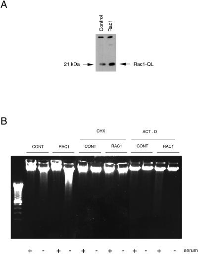
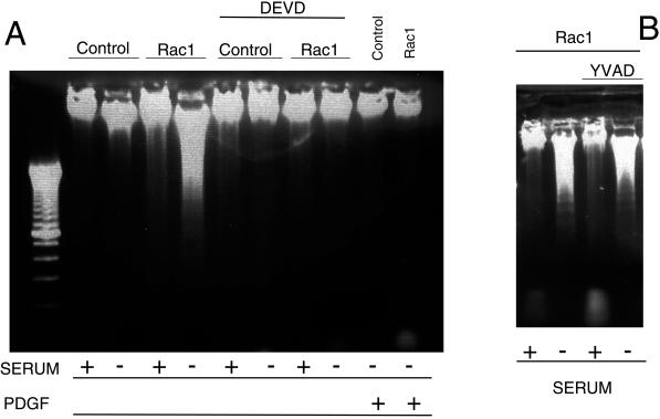
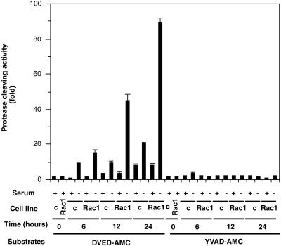
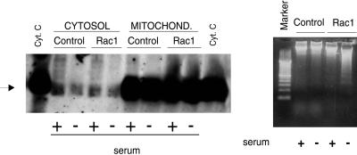
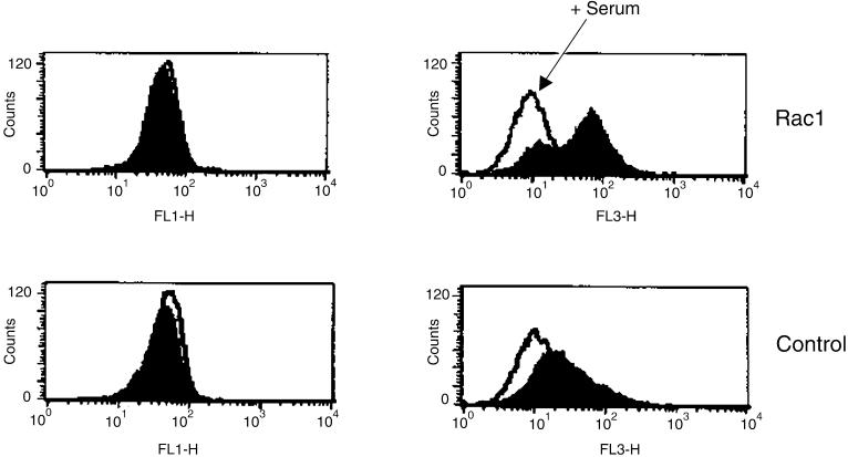
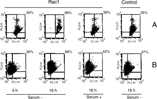
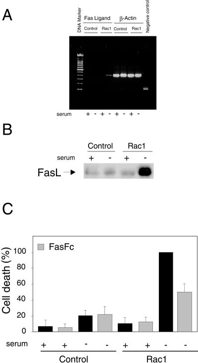
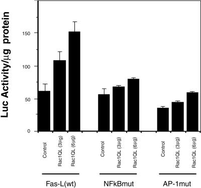
Rho proteins, members of the Ras superfamily of GTPases, are critical elements in signal transduction pathways governing cell proliferation and cell death. Different members of the family of human Rho GTPases, including RhoA, RhoC, and Rac1, participate in the regulation of apoptosis in response to cytokines and serum deprivation in different cell systems. Here, we have characterized the mechanism of apoptosis induced by Rac1 in NIH 3T3 cells. It requires protein synthesis and caspase-3 activity, but it is independent of the release of cytochrome c from mitochondria. Moreover, an increase in mitochondria membrane potential and the production of reactive oxygen species was observed. Rac1-induced apoptosis was related to the simultaneous increase in ceramide production and synthesis of FasL. Generation of FasL may be mediated by transcriptional regulation involving both c-Jun amino terminal kinase as well as nuclear factor-kappa B-dependent signals. None of these signals, ceramides or FasL, was sufficient to induce apoptosis in the parental cell line, NIH 3T3 cells. However, any of them was sufficient to induce apoptosis in the Rac1-expressing cells. Finally, inhibition of FasL signaling drastically reduced apoptosis by Rac1. Thus, Rac1 seems to induce apoptosis by a complex mechanism involving the generation of ceramides and the de novo synthesis of FasL. These results suggest that apoptosis mediated by Rac1 results from a signaling mechanism that involves biochemical and transcriptional events under control of Rac1.


Figures

Figure 1
Figure 1
Induction of apoptosis by overexpression of the human Rac protein needs mRNA and proteins synthesis. (A) NIH 3T3 cells were transfected with the appropriate plasmids carrying the human gene rac-1 activated by a Leu 61 mutation (QL mutant). Pools of transfected cells were selected for geneticin resistance and equivalent amounts of protein lysates were analyzed by Western blot for the level of expression of the protein by using a specific antibody. Control indicates extracts from cells transfected with the empty vector, pLNCX. Rac1 indicates the constitutively active pLNCX-Rac1 QL mutant. The arrow on the right indicates the position of Rac1QL protein. (B) Analysis of apoptosis by the DNA fragmentation assay after serum deprivation. Cells were incubated for 24 h in DMEM with or without serum and in presence of 50 μM cycloheximide (CHX), actinomycin D (ACT. D). Gel shows the DNA ladder after staining with ethidium bromide.
Figure 2
Figure 2
Effect of caspase protease inhibitors on Rac-induced apoptosis. Rac1 and control NIH 3T3 cells were incubated in either DMEM supplemented with 10% NCS (+) or DMEM without serum (−). The caspase-3 inhibitor acetyl-DEVD-CHO (50 μM) (A) or the caspase-1 inhibitor acetyl-YVAD-CHO (100 μM) (B) was added and cells incubated for an additional period of 24 h. After this period, cells were analyzed for induction of apoptosis by the DNA fragmentation analysis as described in MATERIALS AND METHODS. Where indicated, DMEM supplemented with PDGF (100 nM) was used. Control, NIH 3T3 cells; Rac1, NIH 3T3 cells overexpressing the mutated Rac1-QL protein.
Figure 3
Figure 3
Caspase-3 activation in apoptosis induced by Rac1. NIH 3T3 cells constitutively expressing the Rac1QL protein and cells transfected with the empty plasmid (c) were cultivated for 24 h in DMEM supplemented with 10% new born calf serum (+) or DMEM without serum (−). The cells were collected at 0, 6, 12, and 24 h after treatment and cytoplasmic extracts were tested for protease activity by using specific substrates as indicated in MATERIALS AND METHODS. Caspase-1 and caspase-3 enzyme activity is expressed as fold activation over basal levels. Data are representative of four experiments performed in triplicates, with similar results. Bars represent SD.
Figure 4
Figure 4
Rac1-induced apoptosis progress in the absence of cytochrome c accumulation in the cytosol. (Left) NIH 3T3 cells (Control) and Rac1-expressing cells (Rac1) were incubated in DMEM medium with (+) or without (−) serum for 24 h. After that, cells extracts were obtained and processed for cytochrome c analysis. Both cytosolic (Cytosol) and mitochondrial preparations (Mitochond.) were tested for the presence of cytochrome c by Western blot analysis, by using a monoclonal antibody against cytochrome c. Cyt. C indicates purified cytochrome c. The arrow indicates the position of the cytochrome c. (Right) Parallel cultures of NIH 3T3 cells (Control) and Rac1-expressing cells (Rac1) were incubated in DMEM medium with (+) or without (−) serum for 24 h cells and then analyzed for induction of apoptosis by the DNA fragmentation analysis as described in MATERIALS AND METHODS.
Figure 5
Figure 5
Rac1 induces mitochondrial hyperpolarization. Rac1-transformed cells and NIH 3T3 cells were cultured in the presence or absence of serum during 4 h. Changes in ΔΨm were measured by using JC-1 uptake by flow cytometry in both green (FL-1) and red (Fl-3) channels. White peaks represent JC-1 uptake in the presence of serum (arrow), whereas colored peaks indicate JC-1 stained cells under serum deprivation conditions. A representative result of three experiments is shown.
Figure 6
Figure 6
Rac1 increases both Δψ and ROS production. (A) Analysis of Δψ alterations by DioC6 (FL-1) and cellular death by PI (FL-3) at indicated times. (B) Relationship between ROS production and changes in Δψ. ROS were detected by using HE (FL-3). Two representative results of four experiments are shown.
Figure 7
Figure 7
FasL is generated in Rac1-expressing cells after serum deprivation and it is necessary for apoptosis. (A) NIH 3T3 cells stably expressing Ras-1QL protein (Rac1) or control NIH 3T3 cells (Control) were incubated in DMEM with (+) or without (−) serum for 24 h. Then, cells were processed for FasL expression determined by RT-PCR as described in MATERIALS AND METHODS. FasL expression in shown on the left (Fas Ligand), and as control, the β-actin expression (β-Actin) was determined with the same samples in parallel reactions. Negative control refers to an RT-PCR amplification without primers. (B) Western blot analysis of FasL levels in NIH 3T3 and Rac1 cells in the presence and absence of serum. (C) Cells were incubated with FasFc (0.2 μg/ml) either in control medium or without serum for 24 h. Cell death was estimated by flow cytometry (Annexin V) and expressed as percentage of the value of Rac1 without serum for each experiment (cell death range: 22–38%). Data are the mean ± SD from three independent experiments.
Figure 8
Figure 8
Rac1QL activates transcription of the hFasL promoter through the κB and AP-1 sites. NIH 3T3 fibroblasts were cotransfected with increasing amounts of Rac1QL together with 3 μg of the luciferase reporter constructs containing a 0.9-kb fragment of the human Fas L promoter (left), a mutated form deficient in NF-κB binding (middle), and a mutated form deficient in AP-1 binding (right). Luciferase activity was determined 24 h after transfection. Results are expressed as luciferase activity per milligram of protein extracts. Overexpression of Rac1QL in NIH 3T3 cells induces transcriptional activation of the FasL promoter in a dose-dependent manner (left). Transcriptional regulation of the hFasL promoter by Rac1QL is dependent on functional κB (middle) and AP-1 (right) sites. Data represent the mean of a single experiment performed in triplicate ± SD. Same results were observed in three independent experiments.
Figure 9
Figure 9
Induction of apoptosis by exogenous addition of FasL and/or synthetic ceramides. Analysis of DNA fragmentation was performed essentially as described in MATERIALS AND METHODS. Control, NIH 3T3 cells, and NIH 3T3 overexpressing Rac1 were incubated in DMEM supplemented with 10% NCS until they reached 70% confluence. At this time, cells were incubated in DMEM with (+) or without (−) serum, and treated as follows. (A) Cells were incubated with Fas ligand (50 ng/ml) in the absence or presence of serum for 19 h. (B) Cells were incubated with C2 ceramide (100 μM) in the presence or absence of serum for 48 h. (C) Cells were incubated with Fas ligand (50 ng/ml) plus ceramides C2 (100 μM) in the presence or absence of 10% NCS for 24 h.

References

    1. Ashkenazi A, Dixit VM. Apoptosis control by death and decoy receptors. Curr Opin Cell Biol. 1999;11:255–260. - PubMed
    1. Basu S, Kolesnick R. Stress signals for apoptosis, ceramide and c-Jun kinase. Oncogene. 1998;17:3277–3285. - PubMed
    1. Bazenet CE, Mota MA, Rubin LL. The small GTP-binding protein cdc42 is required for nerve growth factor withdrawal-induced neuronal death. Proc Natl Acad Sci USA. 1998;95:3984–3989. - PMC - PubMed
    1. Bazzoni F, Beutler B. The tumor necrosis factor ligand and receptor families. N Engl J Med. 1996;334:1717–1725. - PubMed
    1. Boldin MP, Mett IL, Varfolomeev EE, Chumakow Y, Shemer-Avin Y, Camonis JH, Wallach D. Self-association of the “death domains” of the p55 tumor necrosis factor (TNF) receptor and Fas/APO1 prompts signaling for TNF and Fas/APO1 effects. J Biol Chem. 1995;270:387–391. - PubMed

Publication types

MeSH terms