

Review
doi: 10.1186/gb-2000-1-1-reviews001.
Epub 2000 Jun 9.
An overview of the structures of protein-DNA complexes
Affiliations
- PMID: 11104519
- PMCID: PMC138832
- DOI: 10.1186/gb-2000-1-1-reviews001
Item in Clipboard
Review
An overview of the structures of protein-DNA complexes
Genome Biol.
2000.
Abstract
On the basis of a structural analysis of 240 protein-DNA complexes contained in the Protein Data Bank (PDB), we have classified the DNA-binding proteins involved into eight different structural/functional groups, which are further classified into 54 structural families. Here we present this classification and review the functions, structures and binding interactions of these protein-DNA complexes.
Figures

Flow diagram showing the selection of the protein-DNA complexes from the PDB (04/01/00). The protein-DNA complexes were grouped into structurally related families using the secondary structure alignment program SSAP (see text).

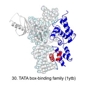
Group I, HTH proteins. The DNA-binding motif is red. The protein binds as a dimer; one monomer is colored blue and the other yellow. The DNA is shown as a space filling model. Family names and numbers are as listed in Table 2; PDB codes are bracketed.

Group II, zinc-coordinating proteins. Colors, numbers and names are as in Figure 1.
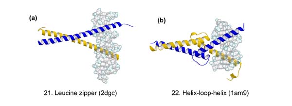
Group III, zipper-type proteins. Colors, numbers and names are as in Figure 1.

Group IV, 'other α helix proteins'. Colors, numbers and names are as in Figure 1.
Group V, β-sheet proteins. Colors, numbers and names are as in Figure 1.

Group VI, the β-hairpin/ribbon proteins. Colors, numbers and names are as in Figure 1.
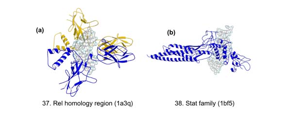
Group VII, 'other DNA-binding proteins'. Colors, numbers and names are as in Figure 1.

Group VIII, the enzymes. Colors, numbers and names are as in Figure 1.
References
-
- Harrison SC. A structural taxonomy of DNA-binding domains. . Nature. 1991;353:715–719. - PubMed
-
- Luisi BF. DNA-protein interaction at high resolution. In DNA-Protein Structural Interactions Edited by Lilley DMJ New York: Oxford University Press, 1995:1–48.
-
- Frishman D, Mewes H-W. PEDANTic genome analysis. Trends Genet. 1997;13:415–416.
-
- Bernstein FC, Koetzler TF, Williams GJB, Meyer EF, Brice MD, Rogers JR, Kennard O, Shimanouchi T, Tasumi M. The Protein Data Bank: a computer-based achival file for macromolecular structures. J Mol Biol. 1977;112:535–542. - PubMed
Publication types
MeSH terms
Substances
LinkOut - more resources
Full Text Sources
Other Literature Sources
