. 2000 Dec;106(11):1351-9.
doi: 10.1172/JCI11093.

An abnormal Ca(2+) response in mutant sarcomere protein-mediated familial hypertrophic cardiomyopathy

Affiliations

An abnormal Ca(2+) response in mutant sarcomere protein-mediated familial hypertrophic cardiomyopathy

D Fatkin et al. J Clin Invest. 2000 Dec.

Abstract

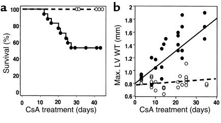
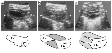
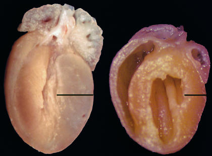
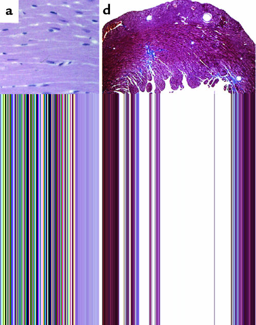
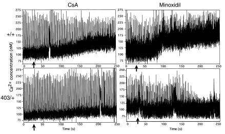
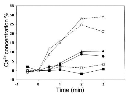
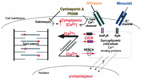
Dominant-negative sarcomere protein gene mutations cause familial hypertrophic cardiomyopathy (FHC), a disease characterized by left-ventricular hypertrophy, angina, and dyspnea that can result in sudden death. We report here that a murine model of FHC bearing a cardiac myosin heavy-chain gene missense mutation (alphaMHC(403/+)), when treated with calcineurin inhibitors or a K(+)-channel agonist, developed accentuated hypertrophy, worsened histopathology, and was at risk for early death. Despite distinct pharmacologic targets, each agent augmented diastolic Ca(2+) concentrations in wild-type cardiac myocytes; alphaMHC(403/+) myocytes failed to respond. Pretreatment with a Ca(2+)-channel antagonist abrogated diastolic Ca(2+) changes in wild-type myocytes and prevented the exaggerated hypertrophic response of treated alphaMHC(403/+) mice. We conclude that FHC-causing sarcomere protein gene mutations cause abnormal Ca(2+) responses that initiate a hypertrophic response. These data define an important Ca(2+)-dependent step in the pathway by which mutant sarcomere proteins trigger myocyte growth and remodel the heart, provide definitive evidence that environment influences progression of FHC, and suggest a rational therapeutic approach to this prevalent human disease.


Figures

Figure 1
Figure 1
(a) Survival of 13 CsA-treated wild-type (open circles) and 18 CsA-treated αMHC403/+ (filled circles) mice. Two αMHC403/+ mice died on the same day on two occasions. Wild-type mice were sacrificed at variable times to provide control specimens for αMHC403/+ mice. (b) Serial assessment of LV wall thickness (measured by transthoracic echocardiography) of wild-type (open circles) and αMHC403/+ (filled circles) mice treated with CsA. The coefficient of correlation is r = 0.83 for LV wall thickness and duration of treatment of αMHC403/+ mice.
Figure 2
Figure 2
Echocardiographic assessment of CsA-treated and untreated αMHC403/+ hearts. Upper panel: Transthoracic echocardiograms of untreated (a) and CsA-treated αMHC403/+ mice (b, day 7; c, day 15). Lower panel: Cardiac morphology defined by echocardiograms of untreated (a) and CsA-treated αMHC403/+ mice (b, day 7; c, day 15).
Figure 3
Figure 3
Gross morphology of hearts from CsA-treated αMHC403/+ (left) and wild-type (right) mice. The αMHC403/+ specimen exhibits marked left- and right-ventricular hypertrophy with cavity obliteration after 30 days of CsA treatment.
Figure 4
Figure 4
Cardiac histopathology in hypertrophic and normal mice. H&E-stained sections from CsA-treated wild-type mice (a) appear normal. Myocyte hypertrophy and disarray is mild in sections (×40 objective lens) from untreated αMHC403/+ mice (b), but marked in sections from CsA-treated αMHC403/+ mice (c). Masson trichrome was used to stain fibrosis (blue) in cardiac sections (×5 objective lens) from αMHC403/+ mice receiving no treatment (d), CsA plus diltiazem (e), or CsA alone (f).
Figure 5
Figure 5
Ca2+ concentrations, assessed in fura-2–loaded αMHC403/+ and wild-type myocytes, before and after CsA or minoxidil treatment. Isolated myocytes from wild-type (+/+) and αMHC403/+ (403/+) mice have comparable calcium concentrations at base line. Addition of CsA (15 μg/ml) or minoxidil (200 μg/ml) (vertical arrows) increases diastolic Ca2+ concentrations (vertical bars) in wild-type, but not mutant, myocytes.
Figure 6
Figure 6
The change (%) in diastolic Ca2+ concentrations in myocytes derived from wild-type (open symbols, dashed lines) or αMHC403/+ (closed symbols, solid lines) mice treated with minoxidil (triangles), CsA (circles), or CsA plus diltiazem (squares). Diltiazem (28 μg/ml) administration began 20 seconds before addition of CsA. Each data point represents the average Ca2+ concentration from ten myocytes. After 2 and 3 minutes of treatment with either CsA or minoxidil, the Ca2+ concentration in wild-type myocytes was significantly different from the Ca2+ concentration in mutant myocytes treated with the same drug (P < 0.02) and was significantly different from the Ca2+ concentration in wild-type myocytes treated with diltiazem and CsA or minoxidil (P < 0.01).
Figure 7
Figure 7
The pathway leading from sarcomere protein gene mutation to hypertrophic cardiomyopathy and the role of an abnormal Ca2+ response. CsA and FK506 may increase cytoplasmic Ca2+ through interaction with calcineurin or by activation of the L-type Ca2+ channel. Cytoplasmic Ca2+ enters the sarcoplasmic reticulum by way of an ATPase-dependent calcium pump (SERCA) and exits by way of the inositol triphosphate receptor (InsP3R) and the ryanodine receptor (RyR). Small increases in Ca2+ trigger Ca2+-induced Ca2+ release (CICR) primarily through the RyR. Considerable Ca2+ is stored in the sarcomere. We hypothesize that sarcomeres containing mutant myosins (denoted by asterisks) store more Ca2+ than normal sarcomeres, causing a reduction in sarcoplasmic reticulum Ca2+ that signals a hypertrophic response. Most Ca2+ in the sarcomere and sarcoplasmic reticulum is bound to carrier proteins, whereas most Ca2+ in the cytoplasm is free. Diltiazem is an inhibitor of the L-type Ca2+ channel, whereas minoxidil is an activator of the K+ channel.

Comment in

  • Making matters worse for a broken heart.
    Frey N, Olson EN. Frey N, et al. J Clin Invest. 2000 Dec;106(12):1437-9. doi: 10.1172/JCI11733. J Clin Invest. 2000. PMID: 11120749 Free PMC article. No abstract available.

References

    1. Maron BJ, et al. Prevalence of hypertrophic cardiomyopathy in a general population of young adults. Echocardiographic analysis of 4111 subjects in the CARDIA Study. Coronary Artery Risk Development in (Young) Adults. Circulation. 1995;92:785–789. - PubMed
    1. Colluci, W.S., and Braunwald, E.B. 1997. Pathophysiology of heart failure. In Heart disease. E. Braunwald, editor. W.B. Saunders Co. Philadelphia, Pennsylvania, USA. 394–420.
    1. Benjamin EJ, Levy D. Why is left ventricular hypertrophy so predictive of morbidity and mortality? Am J Med Sci. 1999;317:168–175. - PubMed
    1. Fatkin, D., Seidman, J.G., and Seidman, C.E. 2000. Hypertrophic cardiomyopathy. In Cardiovascular medicine. J.T. Willerson and J.N. Cohn, editors. W.B. Saunders Co. Philadelphia, Pennsylvania, USA. 1055–1075.
    1. Hunter, J.J., Grace, A., and Chien, K.R. 1999. Molecular and cellular biology of cardiac hypertrophy and failure. In Molecular basis of cardiovascular disease. K.R. Chien, editor. W.B. Saunders Co. Philadelphia, Pennsylvania, USA. 211–250.

Publication types

MeSH terms