. 2000 Dec 4;192(11):1553-62.
doi: 10.1084/jem.192.11.1553.

CXC chemokine receptor 5 expression defines follicular homing T cells with B cell helper function

Affiliations

CXC chemokine receptor 5 expression defines follicular homing T cells with B cell helper function

P Schaerli et al. J Exp Med. .

Abstract

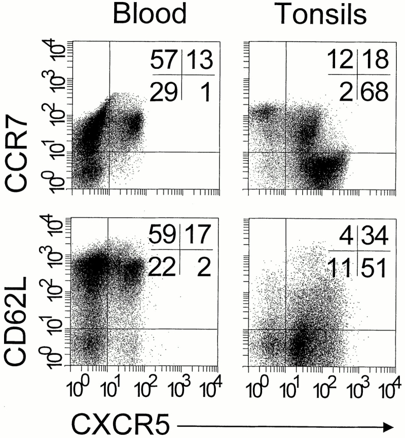
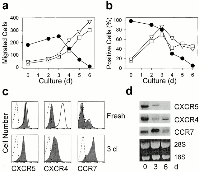
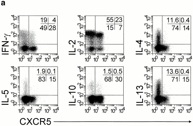
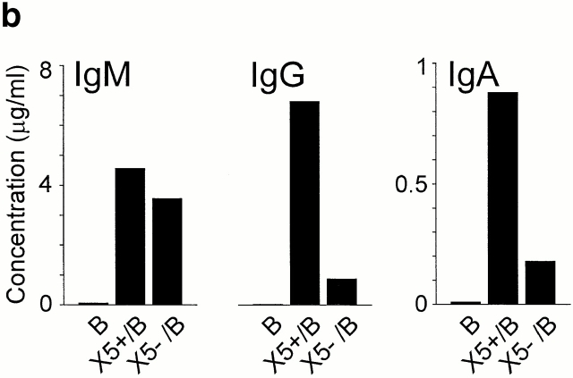
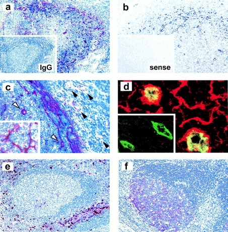
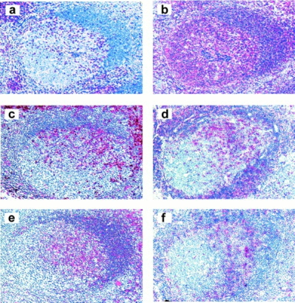
Leukocyte traffic through secondary lymphoid tissues is finely tuned by chemokines. We have studied the functional properties of a human T cell subset marked by the expression of CXC chemokine receptor 5 (CXCR5). Memory but not naive T cells from tonsils are CXCR5(+) and migrate in response to the B cell-attracting chemokine 1 (BCA-1), which is selectively expressed by reticular cells and blood vessels within B cell follicles. Tonsillar CXCR5(+) T cells do not respond to other chemokines present in secondary lymphoid tissues, including secondary lymphoid tissue chemokine (SLC), EBV-induced molecule 1 ligand chemokine (ELC), and stromal cell-derived factor 1 (SDF-1). The involvement of tonsillar CXCR5(+) T cells in humoral immune responses is suggested by their localization in the mantle and light zone germinal centers of B cell follicles and by the concomitant expression of activation and costimulatory markers, including CD69, HLA-DR, and inducible costimulator (ICOS). Peripheral blood CXCR5(+) T cells also belong to the CD4(+) memory T cell subset but, in contrast to tonsillar cells, are in a resting state and migrate weakly to chemokines. CXCR5(+) T cells are very inefficient in the production of cytokines but potently induce antibody production during coculture with B cells. These properties portray CXCR5(+) T cells as a distinct memory T cell subset with B cell helper function, designated here as follicular B helper T cells (T(FH)).


Figures

Figure 1
Figure 1
CCR7 and CD62L are downmodulated on tonsillar CXCR5+ T lymphocytes. Freshly isolated blood and tonsillar lymphocytes were triple stained with antibodies to CD4, CXCR5, and CCR7 or CD62L and analyzed by flow cytometry. Dot plots show cells gated for the expression of CD4. Quadrants were set according to the staining of control antibodies and the percentage of cells in each quadrant is indicated. Comparable results were obtained with five additional blood and tonsillar cell preparations.
Figure 2
Figure 2
Inhibition of migration to BCA-1 and CXCR5 expression during culture of tonsillar CXCR5+ T cells. Freshly isolated tonsillar CXCR5+ T cells were activated with immobilized anti-CD3 for up to 6 d and examined for in vitro migration (a); chemokine receptor expression was assessed by flow cytometry (b and c) and Northern blot analysis (d). (a) Chemotaxis in response to 1,000 nM BCA-1 (•), 100 nM SDF-1 (□), and 100 nM ELC (▿; mean number of migrated cells per 5 HPFs in triplicate wells). The data are representative of five independent experiments. (b) The percentage of cells positive for CXCR5 (•), CXCR4 (□), and CCR7 (▿), with gates set at 99% isotype-matched control antibody staining. (c) Expression of CXCR5, CXCR4, and CCR7 (shaded) in freshly isolated cells and in cells treated with anti-CD3 for 3 d. Broken lines represent the staining of isotype-matched control antibodies, and solid lines show chemokine receptor expression after overnight culture under nonstimulatory conditions. (d) Northern blot analysis of CXCR5, CXCR4, and CCR7 transcripts with total RNA from freshly isolated cells and after treatment with anti-CD3 for 3 and 6 d.
Figure 3
Figure 3
Lack of cytokine production but induction of IgG/IgA synthesis by CXCR5+ T cells. (a) CD4+CD45RO+ T cells were isolated from blood by negative selection. After polyclonal stimulation, CXCR5+ and CXCR5 CD4+ T cells were examined for intracellular production of IFN-γ, IL-2, IL-4, IL-5, IL-10, and IL-13 by three-color flow cytometry. Dot plots show cells gated for CD4. Quadrants were set according to the staining of isotype-matched control antibodies and the percentage of cells in each quadrant is indicated. Results are representative of a total of 11 experiments with memory T cell preparations from different blood and tonsil donors. (b) CXCR5+ T cells induce antibody production in tonsillar B cells. Tonsillar B cells were cultured for 10 d either alone (B) or in the presence of CXCR5+ (X5+/B) or CXCR5 T cells (X5/B), and IgG, IgA, and IgM concentrations in the culture supernatants were determined by ELISA. A representative example out of four independent experiments is shown.
Figure 3
Figure 3
Lack of cytokine production but induction of IgG/IgA synthesis by CXCR5+ T cells. (a) CD4+CD45RO+ T cells were isolated from blood by negative selection. After polyclonal stimulation, CXCR5+ and CXCR5 CD4+ T cells were examined for intracellular production of IFN-γ, IL-2, IL-4, IL-5, IL-10, and IL-13 by three-color flow cytometry. Dot plots show cells gated for CD4. Quadrants were set according to the staining of isotype-matched control antibodies and the percentage of cells in each quadrant is indicated. Results are representative of a total of 11 experiments with memory T cell preparations from different blood and tonsil donors. (b) CXCR5+ T cells induce antibody production in tonsillar B cells. Tonsillar B cells were cultured for 10 d either alone (B) or in the presence of CXCR5+ (X5+/B) or CXCR5 T cells (X5/B), and IgG, IgA, and IgM concentrations in the culture supernatants were determined by ELISA. A representative example out of four independent experiments is shown.
Figure 4
Figure 4
BCA-1 expression in tonsillar follicles. (a and c) Immunostaining of BCA-1 (red) in frozen tonsillar sections; inset in panel a shows control staining with isotype-matched control. (b) In situ hybridization of BCA-1 transcripts using an antisense probe (black) on paraffin sections; no staining is observed with a sense probe (inset in b). Positivity in the mantle zone is associated with reticular cells, as highlighted in the inset of c, and follicular HEVs (white arrowheads in c) but not T zone HEVs (black arrowheads in c). (d) Confocal microscopic analysis of BCA-1 (red) and CD31 (green) expression in follicular HEVs and T zone HEVs (inset in d). Immunostaining of interdigitating dendritic cells with an anti–S-100 antibody (e) and follicular dendritic cells with an anti-CD21 antibody (f) is shown to highlight the nonoverlapping localization of BCA-1–producing reticular cells. Original magnifications: (a, e, and f) ×100; (b and c) ×200; and (d) ×1,600.
Figure 5
Figure 5
Phenotypic characterization of follicular T cells. Immunostaining (red) on paraffin sections is shown for CD3 (a), CD20 (b), CD45RO (c), and HLA-DR (e), and on frozen sections for CD69 (d) and ICOS (f). Original magnification: ×100.

Comment in

Similar articles

Cited by

References

    1. Baggiolini M. Chemokines and leukocyte traffic. Nature. 1998;392:565–568. - PubMed
    1. Loetscher P., Moser B., Baggiolini M. Chemokines and their receptors in lymphocyte traffic and HIV infection. Adv. Immunol. 2000;74:127–180. - PubMed
    1. Murphy P.M., Baggiolini M., Charo I.F., Hebert C.A., Horuk R., Matsushima K., Miller L.H., Oppenheim J.J., Power C.A. International union of pharmacology. XXII. Nomenclature for chemokine receptors. Pharmacol. Rev. 2000;52:145–176. - PubMed
    1. Sallusto F., Mackay C.R., Lanzavecchia A. The role of chemokine receptors in primary, effector, and memory immune responses. Annu. Rev. Immunol. 2000;18:593–620. - PubMed
    1. Zlotnik A., Yoshie O. Chemokinesa new classification system and their role in immunity. Immunity. 2000;12:121–127. - PubMed

Publication types

MeSH terms