. 2000 Dec 19;97(26):14461-6.
doi: 10.1073/pnas.250491597.

Maternal germ-line transmission of mutant mtDNAs from embryonic stem cell-derived chimeric mice

Affiliations

Maternal germ-line transmission of mutant mtDNAs from embryonic stem cell-derived chimeric mice

J E Sligh et al. Proc Natl Acad Sci U S A. .

Abstract

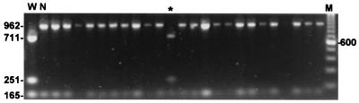
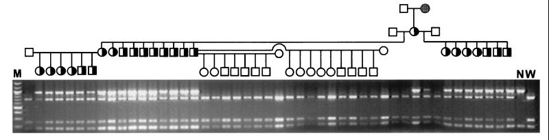
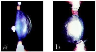
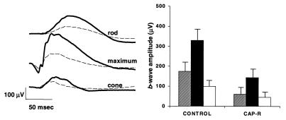
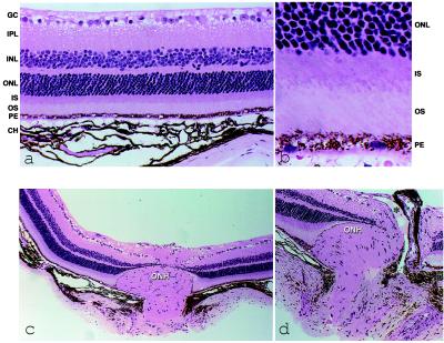
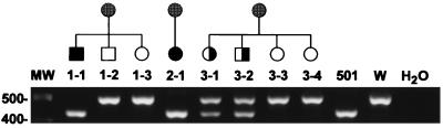
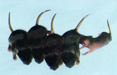
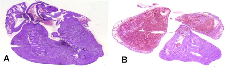
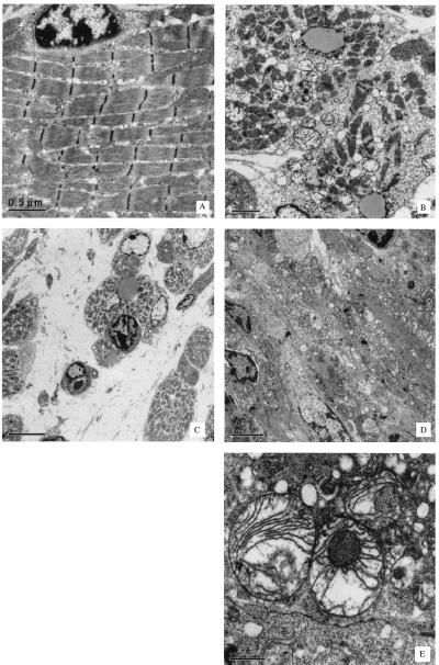
We report a method for introducing mtDNA mutations into the mouse female germ line by means of embryonic stem (ES) cell cybrids. Mitochondria were recovered from the brain of a NZB mouse by fusion of synaptosomes to a mtDNA-deficient (rho degrees ) cell line. These cybrids were enucleated and the cytoplasts were electrofused to rhodamine-6G (R-6G)-treated female ES cells. The resulting ES cell cybrids permitted transmission of the NZB mtDNAs through the mouse maternal lineage for three generations. Similarly, mtDNAs from a partially respiratory-deficient chloramphenicol-resistant (CAP(R)) cell line also were introduced into female chimeric mice and were transmitted to the progeny. CAP(R) chimeric mice developed a variety of ocular abnormalities, including congenital cataracts, decreased retinal function, and hamaratomas of the optic nerve. The germ-line transmission of the CAP(R) mutation resulted in animals with growth retardation, myopathy, dilated cardiomyopathy, and perinatal or in utero lethality. Skeletal and heart muscle mitochondria of the CAP(R) mice were enlarged and atypical with inclusions. This mouse ES cell-cybrid approach now provides the means to generate a wide variety of mouse models of mitochondrial disease.


Figures

Figure 1
Figure 1
Genotyping of individual CC9.3.1 (mtNZB) ES cell cybrid clones for NZB mtDNA content using a BamHI mtDNA polymorphism at np 4277. N is the NZB mtDNA. W is the common haplotype (wild type) mtDNA from LM(TK), the parent cell line of LMEB4. M indicates molecular weight size standards. With the exception of the clone in the lane marked *, the common haplotype was not detected.
Figure 2
Figure 2
Introduction of the NZB mtDNA into the female mouse germ line and its maternal inheritance. DNA isolated from tail biopsy tissue was PCR-amplified and digested. The highly chimeric female is indicated by the checkered circle on the top line of the pedigree. She was mated to a B6 male to generate the founding female. Heteroplasmic individuals are represented by half-shaded squares (males) or circles (females). B6 mice were used as mates to test the inheritance of the NZB mtDNA. W is the common haplotype (wild type) mtDNA from the 129SvEv ES cell line CC9.3.1. N is the NZB mtDNA.
Figure 3
Figure 3
Slit lamp biomicroscopic examination of CAPR chimera mouse lens showing cataracts. (A) The normal clear lens of a control 129S4 mouse. (B) A fetal nuclear cataract of a CAPR chimera.
Figure 4
Figure 4
Electroretinograms of CAPR chimeric mice. (A) Representative responses of rods (Top), mixed rods and cones (Middle), and cones (Bottom) for wild-type 129S4 (solid line) and CAPR chimeric (dashed line) animals. (B) The mean maximum b-wave amplitudes with standard deviation bars for rods (cross hatched bars), mixed rods and cones (filled bars), and cones (open bars) for three representative 129S4 and CAPR chimeric mice.
Figure 5
Figure 5
Retinal light histology of 6-mo-old CAPR chimeric mice. (A) Preservation of retinal layers and a gliotic membrane on the inner retina (Magnified ×400.) (B) The pigment epithelial vacuolization and full preservation of outer photoreceptors. (Magnified ×600.) (C) The optic nervehead (ONH) light histology demonstrating hamartomatous-like changes intraocularly, with a gliotic membrane emanating from the ONH surface. (Magnified ×200.) (D) A similar ONH finding but with the presence of an optic pit (*; magnified ×300) in another CAPR chimeric mouse. GC = ganglion cell layer, IPL = inner plexiform layer, INL = inner nuclear layer, ONL = outer nuclear layer, IS = inner segment, OS = outer segment of photoreceptors, and PE = pigment epithelium.
Figure 6
Figure 6
Germ-line transmission of CAPR mtDNAs into newborn mice. Chimeric females were mated with B6 males, and tissue DNAs were extracted from offspring. The mtDNA 16S rRNA gene was PCR-amplified and the 601-bp PCR product was digested with TaiI. A 434-bp fragment is indicative of CAPR mtDNAs, whereas a 502-bp fragment size is associated with wild-type mtDNA. MW indicates the molecular weight marker. 1-1, 1–2, and 1–3 are from the first transmitting litter. 2-1 is a pup from a second chimeric female's litter. 3-1, 3-2, 3-3, and 3–4 are from a third chimeric female's litter. 501 is the 501-1 cell line containing CAPR mtDNA. W is the wild-type mtDNA from the LM(TK) cell line.
Figure 7
Figure 7
Growth retardation of an agouti mouse harboring CAPR mtDNA. The 10-day-old agouti pup is shown with four black littermates.
Figure 8
Figure 8
Dilated cardiomyopathy in a CAPR neonate on the first day of life. (A) A wild-type B6,129S4 control heart. (B) The CAPR neonatal heart.
Figure 9
Figure 9
Ultrastructural abnormalities of CAPR mouse skeletal muscle and heart. (A–C) Skeletal muscle samples. (D and E) Heart. (A) Wild-type control skeletal muscle at ×4,400. (B–E) CAPR mutant specimens, B at ×3,400, C at ×1,100, D at ×1,950, and E at ×10,500.

Comment in

Similar articles

Cited by

References

    1. Li Y, Huang T T, Carlson E J, Melov S, Ursell P C, Olson J L, Noble L J, Yoshimura M P, Berger C, Chan P H, et al. Nat Genet. 1995;11:376–381. - PubMed
    1. Graham B, Waymire K, Cottrell B, Trounce I A, MacGregor G R, Wallace D C. Nat Genet. 1997;16:226–234. - PubMed
    1. Melov S, Schneider J A, Day B J, Hinerfeld D, Coskun P, Mirra S S, Crapo J D, Wallace D C. Nat Genet. 1998;18:159–163. - PubMed
    1. Melov S, Coskun P, Patel M, Tunistra R, Cottrell B, Jun A S, Zastawny T H, Dizdaroglu M, Goodman S I, Huang T, et al. Proc Natl Acad Sci USA. 1999;96:846–851. - PMC - PubMed
    1. Esposito L A, Kokoszka J E, Waymire K G, Cottrell B, MacGregor G R, Wallace D C. Free Radical Biol Med. 2000;28:754–766. - PMC - PubMed

Publication types