

doi: 10.1073/pnas.260496497.
Species-specific polyamines from diatoms control silica morphology
Affiliations
- PMID: 11106386
- PMCID: PMC18883
- DOI: 10.1073/pnas.260496497
Item in Clipboard
Species-specific polyamines from diatoms control silica morphology
Proc Natl Acad Sci U S A.
.
Abstract
Biomineralizing organisms use organic molecules to generate species-specific mineral patterns. Here, we describe the chemical structure of long-chain polyamines (up to 20 repeated units), which represent the main organic constituent of diatom biosilica. These substances are the longest polyamine chains found in nature and induce rapid silica precipitation from a silicic acid solution. Each diatom is equipped with a species-specific set of polyamines and silica-precipitating proteins, which are termed silaffins. Different morphologies of precipitating silica can be generated by polyamines of different chain lengths as well as by a synergistic action of long-chain polyamines and silaffins.
Figures
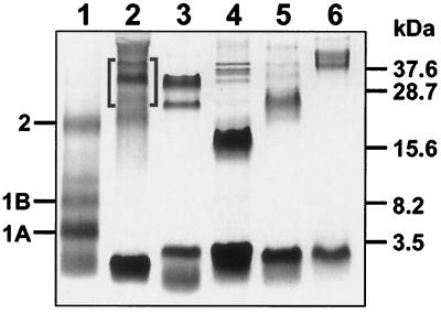
HF extracts from diatom cell walls. Extracts were subjected to
Tris-Tricine SDS/PAGE (11) and stained with Coomassie blue.
Lanes: 1, C. fusiformis (positions of silaffin species
are marked); 2, N. angularis; 3, C.
didymum; 4, C. debilis; 5, E.
zodiacus; 6, S. turris. The brackets in lane 2
mark the three silaffins that were used for silica precipitation (see
Fig. 6).
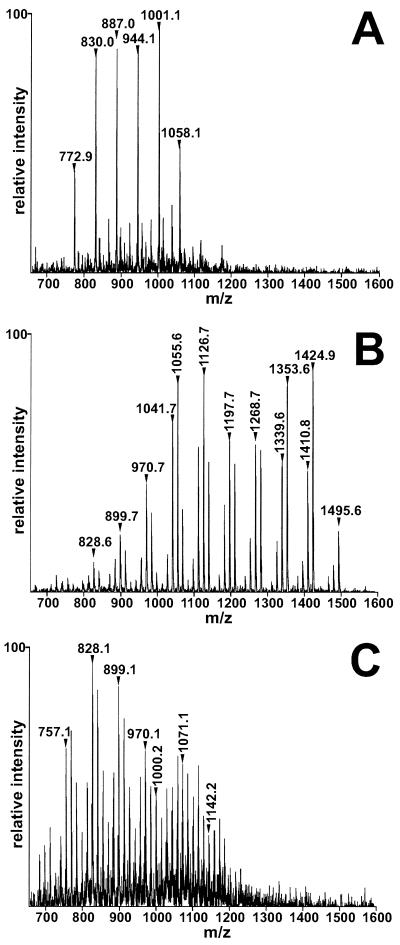
Characterization of diatom polyamines. Electrospray ionization/MS
analysis of purified polyamine fractions. Each peak represents a singly
charged positive ion. Selected peaks are marked by their
m/z units. (A) C.
fusiformis polyamines. (B) C.
didymum polyamines. (C) N.
angularis polyamines.
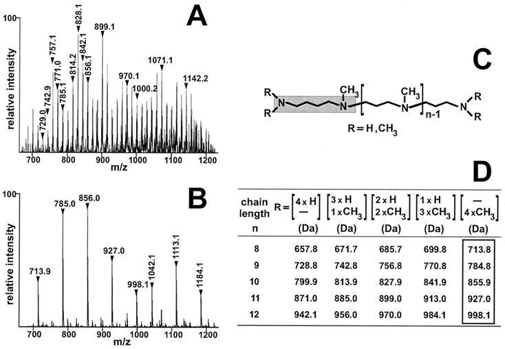
Permethylation analysis of the polyamines from N.
angularis. (A and B) Electrospray
ionization/MS analysis of singly charged positive ions.
(A) Unmodified polyamines from N.
angularis. Two peak series, which are separated by 71 units,
are denoted by vertical numbers that indicate the
m/z value of the corresponding peak.
Within each series, peak masses differ by 14 units. The horizontal
numbers indicate selected molecules whose masses differ by 71 units.
(B) Polyamines from N. angularis after
permethylation. Two peak series can be discerned, which are denoted by
horizontal numbers (putrescine basis) and vertical numbers (ornithine
basis), respectively. Within each series, peak masses differ by 71
units. (C) Scheme of proposed general polyamine
structure. The gray box highlights the putrescine moiety.
(D) Theoretical molecular masses of putrescine-based
polyamine molecules. Each line corresponds to a given polyamine chain
length and lists the theoretical molecular masses of methylation
isoforms. The molecular masses of fully methylated isoforms are
boxed.
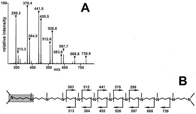
Fragmentation analysis. (A) Product ion spectrum
obtained by collision-induced fragmentation of the (m +
H)+ = 856 ion (see mass signal in Fig.
3B). Two series of ions were detected that differ by 14
units. Within each series, neighboring peaks differ by 71 units.
(B) Proposed structure (schematic) of the (m +
H)+ = 856 ion. Cleavage positions that lead to the
observed fragment ions are depicted by rectangular arrows, and the
corresponding masses are indicated. The putrescine residue is
highlighted by a gray box.
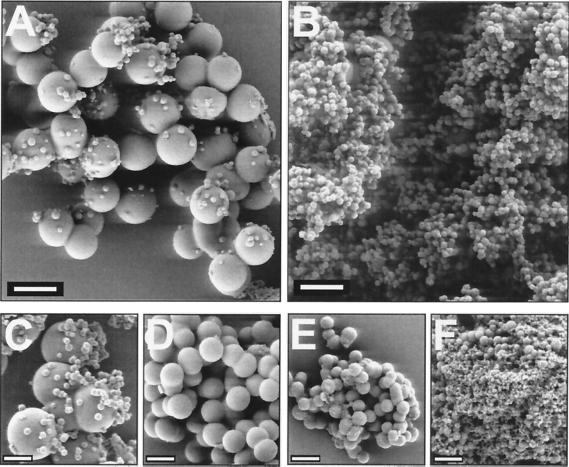
Silica precipitates induced by N. angularis polyamines.
For precipitation, polyamines of molecular masses from 1,000 to 1,250
(A) and 600 to 750 Da (B) were used.
(C–F) The natural mixture of polyamines
(molecular masses 600–1,250 Da) was used for investigating the pH
dependence on polyamine-induced silica precipitation.
(C) pH 5.4. (D) pH 6.3.
(E) pH 7.2. (F) pH 8.3. The polyamine
concentration in each solution was 0.85 mg/ml. [Scale bars, 1 μm
(A and B) and 500 nm
(C–F).]
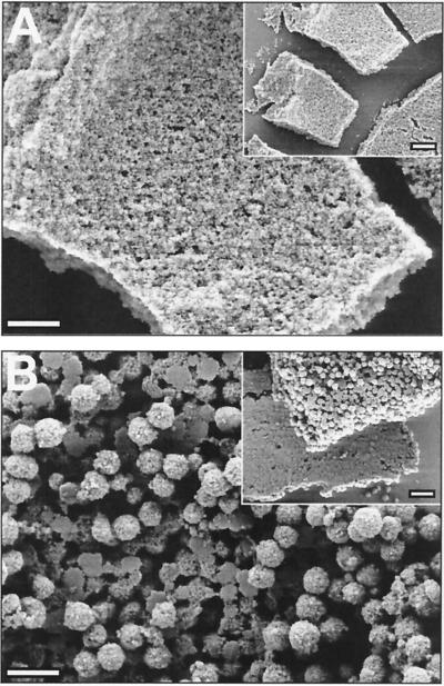
Effect on silica morphology of combining N. angularis
polyamines and silaffins. For precipitation in A,
enriched silaffins (see brackets in Fig. 1, lane 2) at a concentration
of 3 mg/ml were used and in B, a mixture of silaffins
(3 mg/ml) and polyamines (0.85 mg/ml) was used.
(Insets) Larger views of the precipitates. [Scale bars,
500 nm, 1 μm (Insets).]
References
-
- Lowenstam H A. Science. 1981;211:1126–1131. - PubMed
-
- Volcani B E. In: Silicon and Siliceous Structures in Biological Systems. Simpson T L, Volcani B E, editors. New York: Springer; 1981. pp. 157–200.
-
- Pickett-Heaps J, Schmid A M M, Edgar L A. In: Progress in Phycological Research. Round F E, Chapman D J, editors. Vol. 7. Bristol, U.K.: Biopress; 1990. pp. 1–169.
-
- Gordon R, Drum R W. Int Rev Cytol. 1994;150:243–372. - PubMed
-
- Mann S. Nature (London) 1993;365:499–505.
Publication types
MeSH terms
Substances
LinkOut - more resources
Full Text Sources
Other Literature Sources
