. 2000 Dec 5;97(25):13772-7.
doi: 10.1073/pnas.97.25.13772.

A family of peptidoglycan recognition proteins in the fruit fly Drosophila melanogaster

Affiliations

A family of peptidoglycan recognition proteins in the fruit fly Drosophila melanogaster

T Werner et al. Proc Natl Acad Sci U S A. .

Abstract

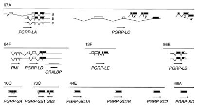
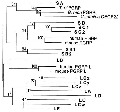
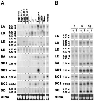
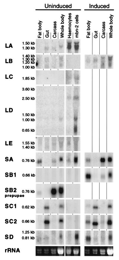
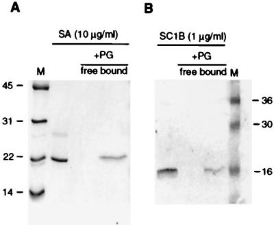
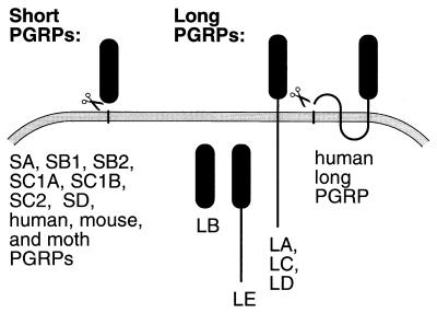
Peptidoglycans from bacterial cell walls trigger immune responses in insects and mammals. A peptidoglycan recognition protein, PGRP, has been cloned from moths as well as vertebrates and has been shown to participate in peptidoglycan-mediated activation of prophenoloxidase in the silk moth. Here we report that Drosophila expresses 12 PGRP genes, distributed in 8 chromosomal loci on the 3 major chromosomes. By analyzing cDNA clones and genomic databases, we grouped them into two classes: PGRP-SA, SB1, SB2, SC1A, SC1B, SC2, and SD, with short transcripts and short 5'-untranslated regions; and PGRP-LA, LB, LC, LD, and LE, with long transcripts and long 5'-untranslated regions. The predicted structures indicate that the first group encodes extracellular proteins and the second group, intracellular and membrane-spanning proteins. Most PGRP genes are expressed in all postembryonic stages. Peptidoglycan injections strongly induce five of the genes. Transcripts from the different PGRP genes were found in immune competent organs such as fat body, gut, and hemocytes. We demonstrate that at least PGRP-SA and SC1B can bind peptidoglycan, and a function in immunity is likely for this family.


Figures

Figure 1
Figure 1
The Drosophila PGRP genes. Maps of PGRP transcripts found at eight chromosomal positions. ORFs are shown as boxes, the PGRP domain in black. Lower boxes are ORFs without PGRP domain. The 5′ region of the PGRP-LD transcript has an ORF related to the human PMI gene, and the antisense strand of the same gene has an ORF similar to cellular retinaldehyde-binding protein (CRALBP). The direction of transcription is indicated by arrows.
Figure 2
Figure 2
Alignment of the PGRP proteins. Amino acids are color coded by chemical properties. Predicted (38) or experimentally determined signal peptides and transmembrane regions (39) are underlined. Intron positions are indicated by arrowheads. Sequences from T. ni, B. mori, C. ethlius, and human are from refs. , , and . The human long PGRP is from chromosome 19 clone CTB-187L3 (AC011492) and the splicing pattern deduced by comparison to the cDNA of the mouse homolog Tagl-α (AF149837).
Figure 3
Figure 3
Phylogenetic tree of the PGRP genes. A maximum parsimony tree was constructed with the amino acid sequences, by using the paup program (41). For branches supported by bootstrap analysis, the percentage of 1,000 replications that support the branch is indicated. Branches supported by at least 50% of the replications are drawn with thick lines.
Figure 4
Figure 4
Expression of the PGRP genes. Northern blot analysis by using 15 μg of total RNA per lane. Identical samples were loaded on several gels and probed for the different genes. Equal loading was checked by ethidium bromide staining; one example is shown (Bottom). Because of crosshybridization between the PGRP-SC1A and SC1B genes, the PGRP-SC1B probe used will detect both transcripts. (A) Developmental expression. L1, L2, L3: first, second, and third instar larvae. (B) Bacterial induction of the PGRP genes. Untreated control flies (c) are compared with flies induced by injection of bacteria (b) of purified peptidoglycan (pg). m, males; f, females.
Figure 5
Figure 5
Tissue distribution of the PGRP gene transcripts. Transcripts are analyzed by Northern blot as in Fig. 4. We also included RNA from hemocytes, isolated from the hemocyte-overproducing mutant l(3)mbn and from the hemocyte-like cell line mbn-2. In each lane, 15 μg of total RNA was loaded. For the samples from fat body, gut, and remaining carcass, we loaded a smaller amount of RNA, corresponding to the same number of larval equivalents as in the whole-body preparation. For PGRP-SB2, we used tissues from prepupae.
Figure 6
Figure 6
Binding of recombinant PGRP-SA and PGRP-SC1B to peptidoglycan from M. luteus. Purified PGRP was incubated with peptidoglycan (PG) at 0.5 mg/ml, and free protein was isolated from bound protein and analyzed on a 15% SDS/PAGE (see Methods). Molecular weights in kilodaltons of the marker proteins (M) are indicated. (A) Binding of recombinant PGRP-SA to peptidoglycan shown with Coomassie brilliant blue staining. (B) Binding of recombinant PGRP-SC1B to peptidoglycan shown by Western blot.
Figure 7
Figure 7
Predicted cellular distribution of the PGRP proteins. All short PGRPs are predicted to be exported after removal of the N-terminal signal peptide. Some of the long PGRPs are likely to be membrane proteins: PGRP-LA, LC, LD, and the human L protein. Others may be retained in the cytoplasm: PGRP-LB and LE. Membrane topology is according to Sipos and von Heijne (39) and signal peptidase cleavage sites according to von Heijne (38).

References

    1. Hultmark D. Nature (London) 1994;367:116–117. - PubMed
    1. Brey P T, Hultmark D. Molecular Mechanisms of Immune Responses in Insects. London: Chapman & Hall; 1998.
    1. Engström Y. Dev Comp Immunol. 1999;23:345–358. - PubMed
    1. Hoffmann J A, Kafatos F C, Janeway C A, Ezekowitz R A B. Science. 1999;284:1313–1318. - PubMed
    1. Ghosh S, May M J, Kopp E B. Annu Rev Immunol. 1998;16:225–260. - PubMed

Publication types