Case Reports
. 2000 Dec 5;97(25):13877-82.
doi: 10.1073/pnas.97.25.13877.

Transplanted fetal striatum in Huntington's disease: phenotypic development and lack of pathology

Affiliations
Case Reports

Transplanted fetal striatum in Huntington's disease: phenotypic development and lack of pathology

T B Freeman et al. Proc Natl Acad Sci U S A. .

Abstract

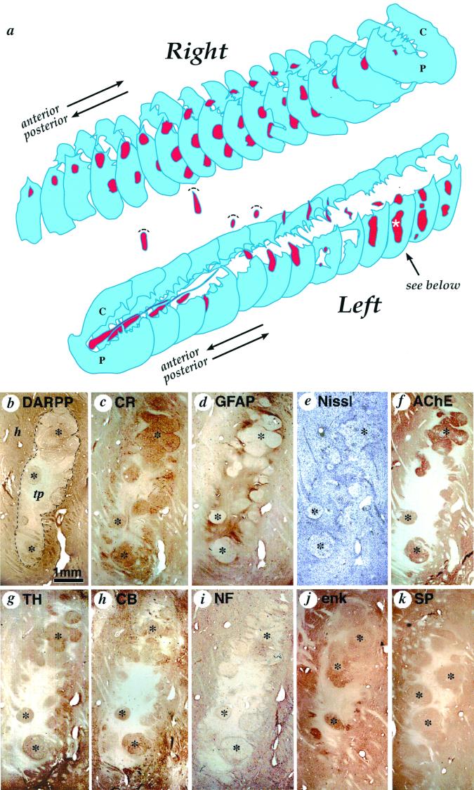
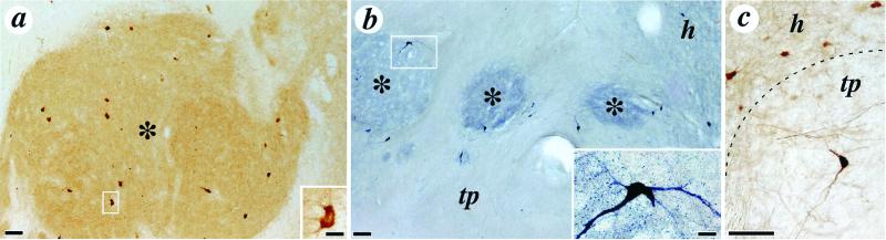
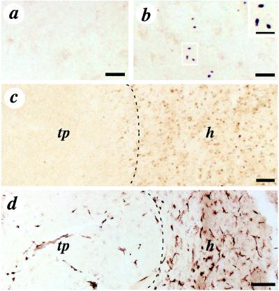
Neural and stem cell transplantation is emerging as a potential treatment for neurodegenerative diseases. Transplantation of specific committed neuroblasts (fetal neurons) to the adult brain provides such scientific exploration of these new potential therapies. Huntington's disease (HD) is a fatal, incurable autosomal dominant (CAG repeat expansion of huntingtin protein) neurodegenerative disorder with primary neuronal pathology within the caudate-putamen (striatum). In a clinical trial of human fetal striatal tissue transplantation, one patient died 18 months after transplantation from cardiovascular disease, and postmortem histological analysis demonstrated surviving transplanted cells with typical morphology of the developing striatum. Selective markers of both striatal projection and interneurons such as dopamine and c-AMP-related phosphoprotein, calretinin, acetylcholinesterase, choline acetyltransferase, tyrosine hydroxylase, calbindin, enkephalin, and substance P showed positive transplant regions clearly innervated by host tyrosine hydroxylase fibers. There was no histological evidence of immune rejection including microglia and macrophages. Notably, neuronal protein aggregates of mutated huntingtin, which is typical HD neuropathology, were not found within the transplanted fetal tissue. Thus, although there is a genetically predetermined process causing neuronal death within the HD striatum, implanted fetal neural cells lacking the mutant HD gene may be able to replace damaged host neurons and reconstitute damaged neuronal connections. This study demonstrates that grafts derived from human fetal striatal tissue can survive, develop, and are unaffected by the disease process, at least for 18 months, after transplantation into a patient with HD.


Figures

Figure 1
Figure 1
(a) Three-dimensional schematic reconstruction of the patient's caudate nucleus and putamen illustrating the sites and relative sizes of bilateral human fetal cell implants as they appeared 18 months after implantation. Caudate nucleus and putamen cross-sections are shown in blue, and transplant tissue is shown in red. Two identified cell implants in the left putamen and three identified transplants in the right putamen were columnar in shape and oriented horizontally following bilateral frontal tracks. The rostral-most left hemisphere tract, intended for the caudate nucleus, was confined to the internal capsule. Volume of this implant was comparatively reduced and more cell dense than any other graft. (b–k) Serial sections through the most posterior graft in the left putamen (indicated by a white asterisk in a), stained with different neurohistological markers to reveal transplant architecture and immunohistochemically stained with antibodies characteristic of striatal neural cell phenotypes: (b) dopamine and cAMP-associated receptor phosphoprotein, (c) calretinin, (d) glial fibrillary acidic protein for astrocytes, (e) cresyl violet (Nissl) stain for general cell perikarya, (f) acetylcholinesterase staining for cholinergic cells and innervation (also see fig. 2a for choline acetyltransferase), (g) tyrosine hydroxylase staining for donor tissue axons, (h) calbindin, (i) 200-kDa human neurofilament (NF-200) for mature axons, (j) enkephalin, and (k) substance P. Cell implant cytoarchitecture (delineated by a dotted line in a) is characterized by two distinct tissue types: roughly spherical zones containing clusters of larger lower density neurons that are acetylcholinesterase-positive (f), glial fibrillary acidic protein-negative (d), and lightly neurofilament-positive (i) (marked by black stars); surrounded by regions of relatively high cell density that are acetylcholinesterase-negative, glial fibrillary acidic protein-negative -positive, and neurofilament-negative. The remaining photomicrographs identify overlapping classes of striatal phenotypes, whereas tyrosine hydroxylase stains host axons that specifically innervate striatal but not nonstriatal tissue. h, host; tp, transplant. (Scale bar = 1 mm.)
Figure 2
Figure 2
Photomicrographs depicting distinctive striatal neuronal phenotypes within the cell implants. (a) Low power photomicrographs illustrating numerous choline acetyltransferase immunoreactive neurons within one striatal graft zone; the morphology of a choline acetyltransferase positive neuron is shown at higher magnification in the Inset. (b) Low-power photomicrograph illustrating nicotinamide adenine dinucleotide phosphate diaphorase immunoreactivity in the same transplant site. The typical morphology of these neurons is illustrated at higher magnification in the Inset. (c) Higher magnification of calbindin-immunoreactive neurons in the host (h) and within the transplant (tp). Striatal zones of the implants are indicated by asterisks in a and b, and the interface of the host and the transplant is delineated by the dotted line in c. (Scale bars: panels, 100 μm; Insets, 20 μm.)
Figure 3
Figure 3
Immunohistochemical staining for the mutant huntingtin protein (EM-48) (a and b), ubiquitin (nuclear inclusion marker) (c), and HLA-DR (microglia and macrophages marker) (d) in the transplant and the host tissue. EM-48 staining of mutant human huntingtin protein, specifically labeling nuclear inclusions, is abundant in the host tissue (b) but not in the transplant (a). Inclusions in b are illustrated at higher magnification in the Inset. (c) Low-power photomicrograph of ubiquitin staining in the host (h) and absence of staining in the transplant (tp) (interface of the transplant and the host indicated by the dotted line). The transplant (left) is clearly devoid of ubiquitin expression. (d) Low-power photomicrographs of HLA-DR staining (interface of the transplant and the host indicated by a dotted line). The transplant contains few HLA-DR positive cells (tp) compared with the host tissue (h), which contains numerous HLA-DR immunoreactive cells. (Scale bars: a and b, 50 μm; Inset, 25 μm; c, 500 μm; d, 100 μm.)

Comment in

References

    1. Dunnett S B. Neuropathol Appl Neurobiol. 1999;25:351–362. - PubMed
    1. Shihabuddin L S, Palmer T D, Gage F H. Mol Med Today. 1999;5:474–480. - PubMed
    1. The Huntington's Disease Collaborative Research Group. Cell. 1993;72:971–983. - PubMed
    1. Ferrante R J, Kowall N W, Richardson E J., Jr J Neurosci. 1991;11:3877–3887. - PMC - PubMed
    1. DiFiglia M, Sapp E, Chase K O, Davies S W, Bates G P, Vonsattel J P, Aronin N. Science. 1997;277:1990–1993. - PubMed

Publication types