. 2000 Dec;157(6):1875-82.
doi: 10.1016/S0002-9440(10)64826-2.

Facilitated wound healing by activation of the Transglutaminase 1 gene

Affiliations

Facilitated wound healing by activation of the Transglutaminase 1 gene

R Inada et al. Am J Pathol. 2000 Dec.

Abstract

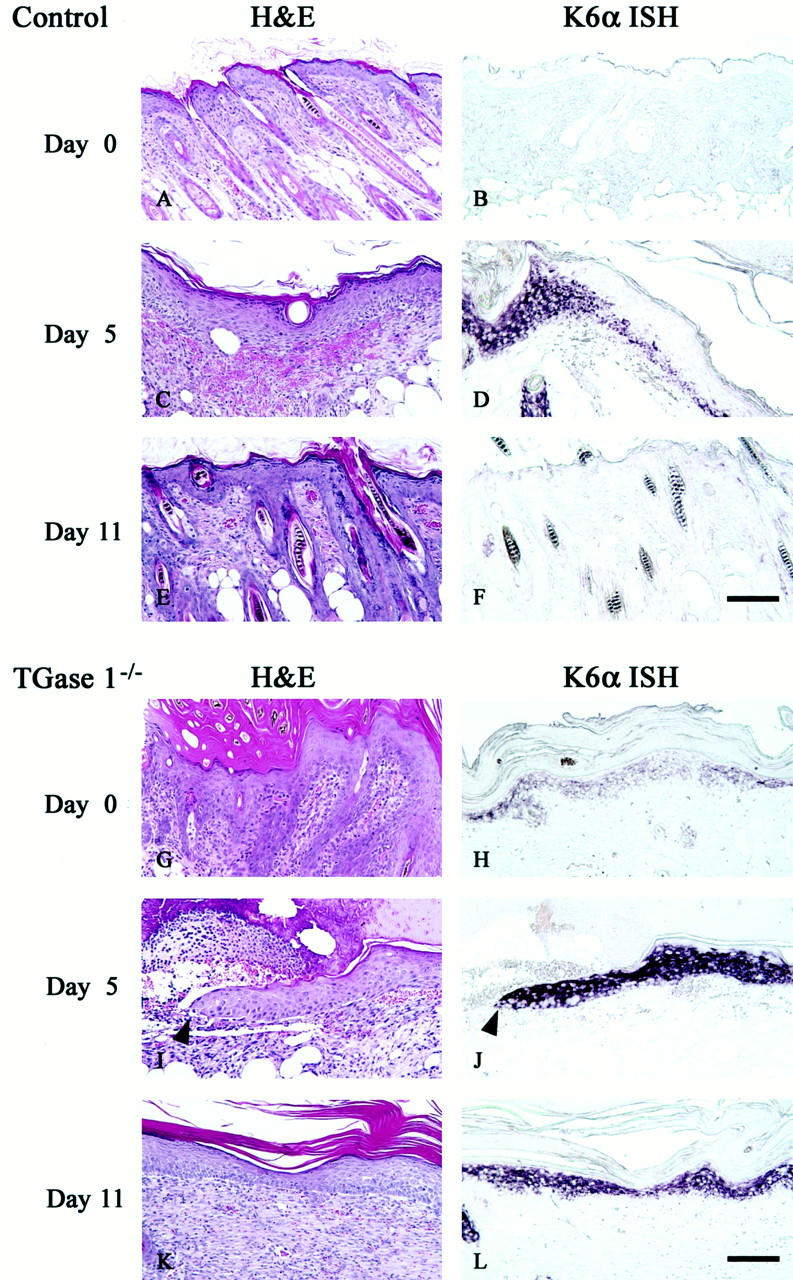
Transglutaminase 1 (TGase 1) is a Ca(2+)-dependent enzyme which catalyzes epsilon-(gamma-glutamyl)lysine cross-linking of substrate proteins such as involucrin and loricrin to generate the cornified envelope at the cell periphery of the stratum corneum. We have shown that disruption of the TGase 1 gene in mice results in neonatal lethality, absence of the cornified envelope, and impaired skin barrier function. Based on the importance of TGase 1 in epidermal morphogenesis, we have now assessed its role in wound healing. In neonatal mouse skin, TGase 1 mRNA as well as keratin 6alpha was induced in the epidermis at the wound edges as early as 2 hours after injury and that expression continued in the migrating epidermis until completion of re-epithelialization. The TGase 1 enzyme co-localized on the plasma membrane of migrating keratinocytes with involucrin, but not with loricrin, which suggests the premature assembly of the cornified envelope. Similar injuries to TGase 1 knockout mouse skins grafted on athymic nude mice showed substantial delays in wound healing concomitant with sustained K6alpha mRNA induction. From these results, we suggest that activation of the TGase 1gene is essential for facilitated repair of skin injury.


Figures

Figure 1.
Figure 1.
Induction of TGase 1 and K6α mRNAs during wound healing in neonatal mouse skin. A 10-mm, full-thickness skin wound was made in BDF1 dorsal skin. The skin was harvested just before (A–C), 2 hours (D–F), 4 hours (G–I), 20 hours (J–L), 48 hours (M–O), and 96 hours (P–R) after the injury. The tissues were stained with H&E (A, D, G, J, M, P), or were subjected to in situ hybridization of TGase 1 (B, E, H, K, N, Q) and K6α mRNA (C, F, I, L, O, R). The induction of TGase 1 and K6α mRNAs begins in the epidermis at the wound edge as early as 2 hours after the injury (E and F). The induction of TGase 1 mRNA becomes more pronounced (H) and is evident even in migrating keratinocytes (K). The induction of TGase 1 mRNA is almost normalized until 96 hours after the injury (Q), at which time K6α mRNA expression is still evident (R). Scale bar, 50 μm.
Figure 2.
Figure 2.
Expression of TGase 1, involucrin, and loricrin in injured neonatal mouse skin. Intact skins (A–C) or injured skins 48 hours after wounding (D–F) were stained with TGase 1 (A and D), involucrin (B and E), or loricrin (C and F) antibodies and were analyzed by confocal fluorescence imaging as described under Materials and Methods. G and H: A higher magnification of D and E showing the migrating edge of keratinocytes expressing TGase 1 and involucrin, respectively. I: A composite image of G over H, showing co-localization of TGase 1 and involucrin at the cell periphery. The arrows show the initial wound sites and the arrowheads indicate the migrating edge of the epithelium. Scale bar, 100 μm (A–F); 25 μm (G–I).
Figure 3.
Figure 3.
Wound healing and induction of K6α mRNA of control and TGase 1−/− skins grafted on nude mice. Skins from control (A–F) and TGase 1−/− neonates (G–L) were grafted onto nude mice. Two weeks after transplantation (A, B, G, H), an injury was made with a 3-mm diameter punch in the center of each graft. Wounded grafts were harvested at 5 (C, D, I, J) and 11 days (E, F, K, L) after the injury. The tissues were stained with H&E (A, C, E, G, I, K), or were subjected to in situ hybridization of K6α mRNA (B, D, F, H, J, L). The control skin grafts mimic adult mouse skin (A) and K6α mRNA is hardly evident (B). The wounds are already closed at day 5 (C), at which time K6α mRNA remains around the initial wound site and is seen only weakly in the regenerated epidermis (D). The control skin grafts are completely remodeled (E) and the K6α mRNA expression subsides (F) 11 days after wounding. On the contrary, the TGase 1−/−-grafted skins show acanthosis with markedly thickened stratum corneum and irregular hair follicles (G), where K6α mRNA is evident (H). At day 5, the wounded TGase 1−/− grafts are covered with a large clot under which epithelial migration is apparently interrupted (I) and K6α mRNA is strongly induced in the migrating epidermis (J). Arrowheads indicate the tip of the migrating epithelium. The re-epithelialization of the TGase 1−/−-grafted skins has been completed at 11 days after wounding (K), but the expression of K6α mRNA is still sustained (L). Scale bar, 100 μm.

References

    1. Folk JE: Transglutaminases. Annu Rev Biochem 1980, 49:517-531 - PubMed
    1. Greenberg CS, Birckbichler PJ, Rice RH: Transglutaminases: multifunctional cross-linking enzymes that stabilize tissues. FASEB J 1991, 5:3071-3077 - PubMed
    1. Singer AJ, Clark RA: Cutaneous wound healing. N Engl J Med 1999, 341:738-746 - PubMed
    1. Bowness JM, Tarr AH, Wong T: Increased transglutaminase activity during skin wound healing in rats. Biochim Biophys Acta 1988, 967:234-240 - PubMed
    1. Muszbek L, Adany R, Mikkola H: Novel aspects of blood coagulation factor XIII. I. Structure, distribution, activation, and function. Crit Rev Clin Lab Sci 1996, 33:357-421 - PubMed

Publication types