. 2000 Dec;157(6):2003-10.
doi: 10.1016/s0002-9440(10)64839-0.

Astroglial expression of human alpha(1)-antichymotrypsin enhances alzheimer-like pathology in amyloid protein precursor transgenic mice

Affiliations

Astroglial expression of human alpha(1)-antichymotrypsin enhances alzheimer-like pathology in amyloid protein precursor transgenic mice

L Mucke et al. Am J Pathol. 2000 Dec.

Abstract

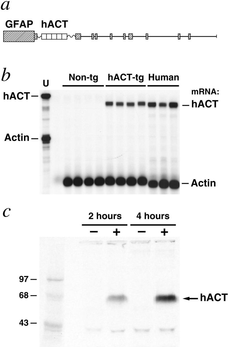
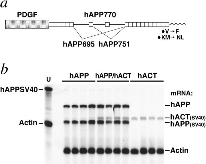
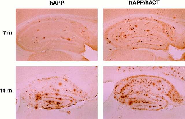
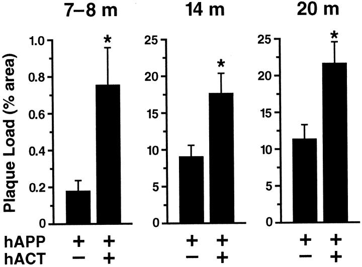
Proteases and their inhibitors play key roles in physiological and pathological processes. Cerebral amyloid plaques are a pathological hallmark of Alzheimer's disease (AD). They contain amyloid-ss (Ass) peptides in tight association with the serine protease inhibitor alpha(1)-antichymotrypsin.(1,2) However, it is unknown whether the increased expression of alpha(1)-antichymotrypsin found in AD brains counteracts or contributes to the disease. We used regulatory sequences of the glial fibrillary acidic protein gene(3) to express human alpha(1)-antichymotrypsin (hACT) in astrocytes of transgenic mice. These mice were crossed with transgenic mice that produce human amyloid protein precursors (hAPP) and Ass in neurons.(4,5) No amyloid plaques were found in transgenic mice expressing hACT alone, whereas hAPP transgenic mice and hAPP/hACT doubly transgenic mice developed typical AD-like amyloid plaques in the hippocampus and neocortex around 6 to 8 months of age. Co-expression of hAPP and hACT significantly increased the plaque burden at 7 to 8, 14, and 20 months. Both hAPP and hAPP/hACT mice showed significant decreases in synaptophysin-immunoreactive presynaptic terminals in the dentate gyrus, compared with nontransgenic littermates. Our results demonstrate that hACT acts as an amyloidogenic co-factor in vivo and suggest that the role of hACT in AD is pathogenic.


Figures

Figure 1.
Figure 1.
Expression of hACT in astrocytes of transgenic mice. a: Astroglial expression of an hACT cDNA was directed by regulatory sequences of a modified murine GFAP gene. SV40 polyadenylation signals at the 3′ end of the hACT cDNA prevent expression of downstream GFAP-coding sequences. Elements are not drawn to scale. b: hACT mRNA levels in brains of humans and transgenic mice. A representative autoradiograph is shown. Total RNA extracted from mouse hemibrains or from the midfrontal gyrus of humans without neurological disease was analyzed by RNase protection assay. The leftmost lane shows signals of undigested radiolabeled riboprobes. The other lanes contained the same riboprobes plus brain RNA (10 μg/lane) from different mice or humans, digested with RNases. Protected mRNA segments are indicated on the right. Non-tg = nontransgenic. Signals were quantitated by phosphorimager analysis: hACT/actin mRNA ratios in hACT mice (0.18, 0.15, 0.17, 0.18) were less variable than those in postmortem human brain tissues (0.18, 0.39, 0.48). c: Production of hACT by transgenic astrocytes. Primary astrocytes were established from transgenic (+) and nontransgenic (−) neonatal mice and metabolically labeled for 2 or 4 hours. hACT was immunoprecipitated with anti-hACT antibodies from conditioned culture medium, separated by sodium dodecyl sulfate-polyacrylamide gel electrophoresis, and detected by autoradiography. Results similar to those shown were obtained with a different hACT antibody. The left lane contains 14C-labeled molecular weight standards.
Figure 2.
Figure 2.
Cerebral transgene expression in hAPP/hACT mice and singly transgenic controls. a: Neuronal expression of an alternatively spliced hAPP minigene was directed by the human PDGF β-chain promoter as described previously. The hAPP minigene expressed in line J9 carries FAD-linked mutations (670/671KM → NL and 717V → F, hAPP770 numbering) that increase the production of Aβ42. Elements are not drawn to scale. b: hACT expression does not alter cerebral hAPP mRNA levels. A representative autoradiograph is shown. Total RNA extracted from mouse hemibrains was analyzed by RNase protection assay. Conventions are as in Figure 1b ▶ . The chimeric hAPPSV40 probe protects human but not mouse amyloid precursor protein; it also protects SV40 sequences in transgene-derived mRNAs. These SV40 sequences provide polyadenylation signals and are of slightly different length in the two transgenes allowing for differentiation of GFAP-hACT-derived (hACT(SV40)) and PDGF-hAPP-derived (hAPP(SV40)) mRNA segments. Signals were quantitated by phosphorimager analysis: singly and doubly transgenic mice (n = 4 per genotype) did not differ significantly in hAPP/actin (0.298 ± 0.053 versus 0.307 ± 0.096) or hACT(SV40)/actin (0.153 ± 0.021 versus 0.160 ± 0.012) mRNA ratios (mean ± SD).
Figure 3.
Figure 3.
Astroglial expression of hACT increases amyloid deposition in hAPP/hACT mice. Brain sections from hAPP mice (left) and hAPP/hACT mice (right) were labeled with the anti-Aβ antibody 3D6 at 7 or 14 months (m) of age. Aβ-immunoreactive deposits in the hippocampus were visualized by immunoperoxidase reaction and light microscopy.
Figure 4.
Figure 4.
Quantitation of plaque load in hAPP and hAPP/hACT mice at different ages. Brain sections of hAPP and hAPP/hACT mice were labeled with the 3D6 antibody at 7 to 8 months (n = 9 per genotype), 14 months (n = 7 per genotype), or 20 months (n = 4 to 6 per genotype) of age. The hippocampal area occupied by Aβ deposits was greater in hAPP/hACT than in hAPP mice at all ages examined. Note the lower scale of the y axis in the youngest age group. Values represent group means ± SEM. *, P < 0.05 versus age-matched hAPP mice (Mann-Whitney U test).
Figure 5.
Figure 5.
Comparable levels of synaptic damage in hAPP and hAPP/hACT mice. The density of SYN-IR presynaptic terminals (a) and the level of GAP-43 immunoreactivity (b) in the molecular layer of the dentate gyrus were determined in nontransgenic controls, singly transgenic mice (hAPP or hACT) and hAPP/hACT doubly transgenic mice (n = 11 to 13 mice per genotype) at 14 to 20 months of age. Data represent group means ± SD. *, P < 0.05 versus nontransgenic controls (Tukey-Kramer test). c: The density of SYN-IR presynaptic terminals did not correlate with the plaque load in hAPP (P = 0.074) or hAPP/hACT (P = 0.88) mice. At 7 to 8 months of age (n = 9 mice per genotype), the density of SYN-IR presynaptic terminals was also similar in hAPP (24.7 ± 1.4) and hAPP/hACT (24.2 ± 1.9) mice and it did not correlate with plaque load (P = 0.76).

References

    1. Abraham CR, Selkoe DJ, Potter H: Immunochemical identification of the serine protease inhibitor α1-antichymotrypsin in the brain amyloid deposits of Alzheimer’s disease. Cell 1988, 52:487-501 - PubMed
    1. Shoji M, Hirai S, Yamaguchi H, Harigaya Y, Ishiguro K, Matsubara E: Alpha1-antichymotrypsin is present in diffuse senile plaques. A comparative study of beta-protein and alpha1-antichymotrypsin immunostaining in the Alzheimer brain. Am J Pathol 1991, 138:247-257 - PMC - PubMed
    1. Johnson WB, Ruppe MD, Rockenstein EM, Price J, Sarthy VP, Verderber LC, Mucke L: Indicator expression directed by regulatory sequences of the glial fibrillary acidic protein (GFAP) gene: in vivo comparison of distinct GFAP-lacZ transgenes. Glia 1995, 13:174-184 - PubMed
    1. Hsia A, Masliah E, McConlogue L, Yu G, Tatsuno G, Hu K, Kholodenko D, Malenka RC, Nicoll RA, Mucke L: Plaque-independent disruption of neural circuits in Alzheimer’s disease mouse models. Proc Natl Acad Sci USA 1999, 96:3228-3233 - PMC - PubMed
    1. Mucke L, Masliah E, Yu G-Q, Mallory M, Rockenstein EM, Tatsuno G, Hu K, Kholodenko D, Johnson-Wood K, McConlogue L: High-level neuronal expression of Aβ1-42 in wild-type human amyloid protein precursor transgenic mice: synaptotoxicity without plaque formation. J Neurosci 2000, 20:4050-4058 - PMC - PubMed

Publication types

MeSH terms

Substances