CSF flow measurement in syringomyelia
- PMID: 11110528
- PMCID: PMC7974308
CSF flow measurement in syringomyelia
Abstract
Background and purpose: CSF circulation has been reported to represent a major factor in the pathophysiology of syringomyelia. Our purpose was to determine the CSF flow patterns in spinal cord cysts and in the subararachnoid space in patients with syringomyelia associated with Chiari I malformation and to evaluate the modifications of the flow resulting from surgery.
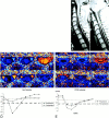
Methods: Eighteen patients with syringomyelia were examined with a 3D Fourier encoding velocity imaging technique. A prospectively gated 2D axial sequence with velocity encoding in the craniocaudal direction in the cervical region was set at a velocity of +/- 10 cm/s. Velocity measurements were performed in the larger portion of the cysts and, at the same cervical level, in the pericystic subarachnoid spaces. All patients underwent a surgical procedure involving dural opening followed by duroplasty. Pre- and postoperative velocity measurements of all patients were taken, with a mean follow-up of 10.2 months. We compared the velocity measurements with the morphology of the cysts and with the clinical data. Spinal subarachnoid spaces of 19 healthy subjects were also studied using the same technique.
Results: A pulsatile flow was observed in syrinx cavities and in the pericystic subarachnoid spaces (PCSS). Preoperative maximum systolic cyst velocities were higher than were diastolic velocities. A systolic velocity peak was well defined in all cases, first in the cyst and then in the PCSS. Higher systolic and diastolic cyst velocities are observed in large cysts and in patients with a poor clinical status. After surgery, a decrease in cyst volume (evaluated on the basis of the extension of the cyst and the compression of the PCSS) was observed in 13 patients. In the postoperative course, we noticed a decrease of systolic and diastolic cyst velocities and a parallel increase of systolic PCSS velocities. Diastolic cyst velocities correlated with the preoperative clinical status of the patients and, after surgery, in patients with a satisfactory foraminal enlargement evaluated on the basis of the visibility of the cisterna magna.
Conclusion: CSF flow measurement constitutes a direct evaluation for the follow-up of patients with syringomyelic cysts. Diastolic and systolic cyst velocities can assist in the evaluation of the efficacy of surgery.
Figures


Comment in
-
Further explanations for the formation of syringomyelia: back to the drawing table.AJNR Am J Neuroradiol. 2000 Nov-Dec;21(10):1778-9. AJNR Am J Neuroradiol. 2000. PMID: 11110524 Free PMC article. No abstract available.
Similar articles
-
Peak systolic and diastolic CSF velocity in the foramen magnum in adult patients with Chiari I malformations and in normal control participants.AJNR Am J Neuroradiol. 2003 Feb;24(2):169-76. AJNR Am J Neuroradiol. 2003. PMID: 12591629 Free PMC article.
-
Elucidating the pathophysiology of syringomyelia.J Neurosurg. 1999 Oct;91(4):553-62. doi: 10.3171/jns.1999.91.4.0553. J Neurosurg. 1999. PMID: 10507374
-
Cerebrospinal fluid flow dynamics study in Chiari I malformation: implications for syrinx formation.Neurosurg Focus. 2000 Mar 15;8(3):E3. doi: 10.3171/foc.2000.8.3.3. Neurosurg Focus. 2000. PMID: 16676926
-
Cerebrospinal fluid flow imaging by using phase-contrast MR technique.Br J Radiol. 2011 Aug;84(1004):758-65. doi: 10.1259/bjr/66206791. Epub 2011 May 17. Br J Radiol. 2011. PMID: 21586507 Free PMC article. Review.
-
Cerebrospinal fluid hydrodynamics in type I Chiari malformation.Neurol Res. 2011 Apr;33(3):247-60. doi: 10.1179/016164111X12962202723805. Neurol Res. 2011. PMID: 21513645 Review.
Cited by
-
Pulsatile cerebrospinal fluid dynamics in Chiari I malformation syringomyelia: Predictive value in posterior fossa decompression and insights into the syringogenesis.J Craniovertebr Junction Spine. 2021 Jan-Mar;12(1):15-25. doi: 10.4103/jcvjs.JCVJS_42_20. Epub 2021 Mar 4. J Craniovertebr Junction Spine. 2021. PMID: 33850377 Free PMC article.
-
Fluid dynamics in syringomyelia cavities: Effects of heart rate, CSF velocity, CSF velocity waveform and craniovertebral decompression.Neuroradiol J. 2018 Oct;31(5):482-489. doi: 10.1177/1971400918795482. Epub 2018 Aug 17. Neuroradiol J. 2018. PMID: 30114970 Free PMC article.
-
Peak systolic and diastolic CSF velocity in the foramen magnum in adult patients with Chiari I malformations and in normal control participants.AJNR Am J Neuroradiol. 2003 Feb;24(2):169-76. AJNR Am J Neuroradiol. 2003. PMID: 12591629 Free PMC article.
-
Characterization of CSF hydrodynamics in the presence and absence of tonsillar ectopia by means of computational flow analysis.AJNR Am J Neuroradiol. 2009 May;30(5):941-6. doi: 10.3174/ajnr.A1489. Epub 2009 Mar 19. AJNR Am J Neuroradiol. 2009. PMID: 19299486 Free PMC article.
-
Prophylactic enlargement of the thecal sac volume by spinal expansion duroplasty in patients with unresectable malignant intramedullary tumors and metastases prior to radiotherapy.Neurosurg Rev. 2020 Feb;43(1):273-279. doi: 10.1007/s10143-018-1051-0. Epub 2018 Nov 14. Neurosurg Rev. 2020. PMID: 30426355
References
-
- Aboulker J. La syringomyélie et les liquides intra-rachidiens. Neurochirurgie 1979;25:1-115 - PubMed
-
- Ball MJ, Dayan AD. Pathogenesis of syringomyelia. Lancet 1972;2:799-801 - PubMed
-
- Williams B. The distending force in the production of “communicating syringomyelia.”. Lancet 1969;2:696-697 - PubMed
-
- Oldfield EH, Muraszko K, Shawker TH, Patronas NJ. Pathophysiology of syringomyelia associated with Chiari I malformation of the cerebellar tonsils: implications for diagnosis and treatment. J Neurosurg 1994;80:3-15 - PubMed
MeSH terms
LinkOut - more resources
Full Text Sources
Medical
