

High-b-value diffusion-weighted MR imaging of adult brain: image contrast and apparent diffusion coefficient map features
- PMID: 11110534
- PMCID: PMC7974278
High-b-value diffusion-weighted MR imaging of adult brain: image contrast and apparent diffusion coefficient map features
Abstract
Background and purpose: Recent improvements in MR gradient technology allow significant increases in diffusion weighting without prohibitive signal-to-noise degradation. The purpose of our investigation was to establish normative references for the signal intensity characteristics and apparent diffusion coefficient values of the adult brain at high b values.
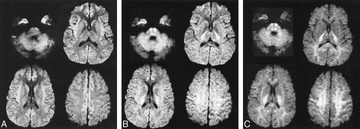
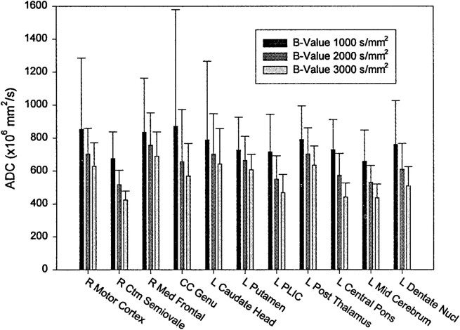
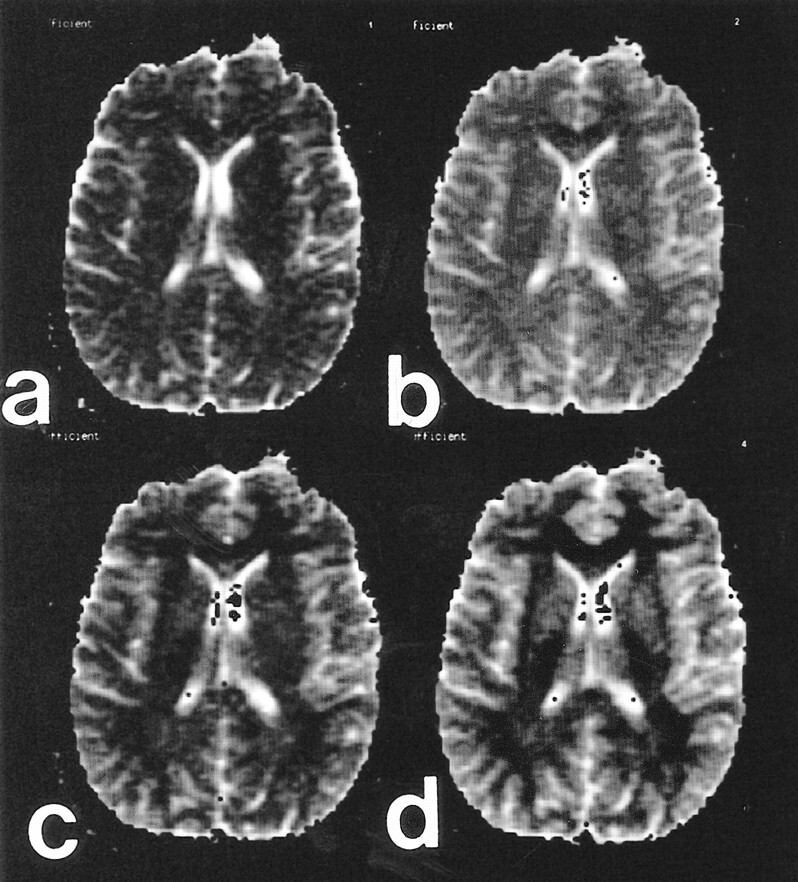
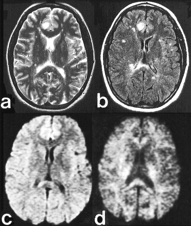
Methods: Fifty adults underwent diffusion-weighted single-shot spin-echo echo-planar MR imaging. Isotropic diffusion-weighted images were obtained with b values of 0, 1,000, 2,000, 2,500, 3,000, and 3,500 s/mm2. Qualitative assessments were made in multiple regions of interest in gray and white matter. Three apparent diffusion coefficient maps were generated for each of six patients with a 2-point technique at a b value of 0 and at b values of 1,000, 2,000, and 3,000 s/mm2.
Results: Increasing b values result in a progressive decrease in the gray to white matter signal intensity ratio. Isointensity between gray and white matter results at b values between 1,000 and 2,000 s/mm2. At b values greater than 2,000, the gray-white pattern reverses relative to the usual b value of 1,000. Apparent diffusion coefficient values were shown to decrease with increasing b values.
Conclusion: Attention to the reversal of gray-white contrast and the dependence of apparent diffusion coefficient on the b value are important in avoiding erroneous assignment of pathologic abnormalities to normal regions. This study provides the normative data for future diffusion investigations performed at high b values.
Figures

Comment in
-
Diffusing into the future.AJNR Am J Neuroradiol. 2000 Nov-Dec;21(10):1780-2. AJNR Am J Neuroradiol. 2000. PMID: 11110526 Free PMC article. No abstract available.
References
-
- Le Bihan D, Breton E, Lallemand D, Grenier P, Cabanis E, Laval-Jeantet M. MR imaging of intravoxel incoherent motions: application to diffusion and perfusion in neurologic disorders. Radiology 1986;161:401-407 - PubMed
-
- Warach S, Chien D, Li W, Ronthal M, Edelman RR. Fast magnetic resonance diffusion-weighted imaging of acute human stroke. Neurology 1992;42:1717-1723 - PubMed
-
- Lovblad KO, Baird AE, Schlaug G, et al. Ischemic lesion volumes in acute stroke by diffusion-weighted magnetic resonance imaging correlate with clinical outcome. Ann Neurol 1997;42:164-170 - PubMed
Publication types
MeSH terms
LinkOut - more resources
Full Text Sources