

Case Reports
Selective neuronal necrosis associated with status epilepticus: MR findings
Affiliations
- PMID: 11110535
- PMCID: PMC7974287
Item in Clipboard
Case Reports
Selective neuronal necrosis associated with status epilepticus: MR findings
AJNR Am J Neuroradiol.
2000 Nov-Dec.
Abstract
We present the MR imaging findings in an autopsy-proven case of selective neuronal necrosis involving the entire left cerebral hemispheric cortex, left thalamus, and contralateral cerebellum following a period of status epilepticus. Imaging findings include diffusion abnormality on diffusion-weighted images and increased intensity on T2-weighted images in the above-mentioned regions of the brain.
Figures
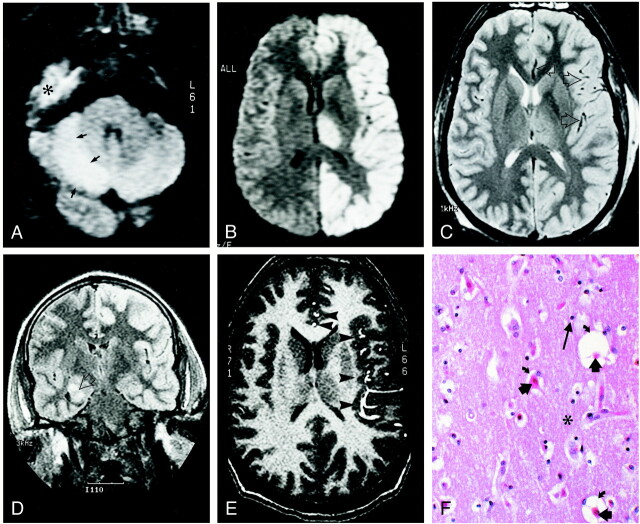
24-year-old man who experienced status epilepticus 4 days prior to imaging. A and B, Diffusion-weighted trace images (9999/91.4 [TR/TE], B = 1000 s/mm2) through the levels of cerebellum (A) and thalami (B) show diffusion abnormality involving the right cerebellar cortex (arrows in A), the entire left hemispheric cortex, and the left thalamus (B). The high signal in the right middle fossa (indicated with * in A) is artifactual. C, Spin-echo T2-weighted (2800/80/1 [TR/TE/excitations]) image through the same level as B shows mildly increased intensity in the diffusely thickened left hemispheric cortex and the left thalamus. Note the relative prominence of signal void in middle and anterior cerebral artery branches (arrows) on the left side. D, Fluid-attenuated inversion recovery coronal image (9002/145/2200 [TR/TE/TI]) reveals the right hippocampal ganglioglioma (arrow). Note increased intensity in the left hemispheric cortex. E, Flow-sensitive gradient-echo T1-weighted image (9.3/2.1/90 degrees [TR/TE/flip angle]). Prominent anterior and middle cerebral artery branches (arrowheads) over the left hemispheric cortex suggest enhanced arterial flow in the left hemisphere. F, Histologic section through the left frontal cortex stained with hemotoxylin and eosin shows eosinophilic neurons (short, thick arrows). The surrounding neuropil (indicated with *) is edematous, as demonstrated by expanded perineural spaces (curved arrows), but there is no fragmentation of the neuropil, and glial cells (long arrow) showed no evidence of necrosis or apoptosis. These findings correspond to selective neuronal necrosis.
Comment in
-
The status of status: seizures are bad for your brain's health.AJNR Am J Neuroradiol. 2000 Nov-Dec;21(10):1782-3. AJNR Am J Neuroradiol. 2000. PMID: 11110527 Free PMC article. No abstract available.
References
-
- Wasterlain CG, Fujikawa DG, Penix LR, Sankar R. Pathophysiological mechanisms of brain damage from status epilepticus. Epilepsia 1993;34:37-153 - PubMed
-
- Fujikawa DG. The temporal evolution of neuronal damage from pilocarpin-induced status epilepticus. Brain Research 1996;725:11-22 - PubMed
-
- Duncan R, Patterson J, Bone I, Wyper DJ. Reversible cerebellar diaschisis in focal epilepsy. Lancet 1987;12:625-626 - PubMed
-
- Fazekas F, Kapeller P, Schmidt R, et al. Magnetic resonance imaging and spectroscopy findings after focal status epilepticus. Epilepsia 1995;36:946-949 - PubMed
-
- Wieshman UC, Symms MR, Shorvon SD. Diffusion changes in status epilepticus. Lancet 1997;350:493-494 - PubMed
Publication types
MeSH terms
LinkOut - more resources
Full Text Sources
Medical