

The choline/creatine ratio in five benign neoplasms: comparison with squamous cell carcinoma by use of in vitro MR spectroscopy
- PMID: 11110549
- PMCID: PMC7974295
The choline/creatine ratio in five benign neoplasms: comparison with squamous cell carcinoma by use of in vitro MR spectroscopy
Abstract
Background and purpose: The choline (Cho)/creatine (Cr) ratio has been shown to be a reliable proton MR spectroscopy metabolic marker for differentiating squamous cell carcinoma (SCCA) from normal muscle in the upper aerodigestive tract. However, it is unclear whether the Cho/Cr ratio can be used to differentiate a malignant tumor from a benign neoplasm in the extracranial head and neck. Our purpose was to determine whether the Cho/Cr ratio can be used to differentiate benign from malignant tumors in this region.
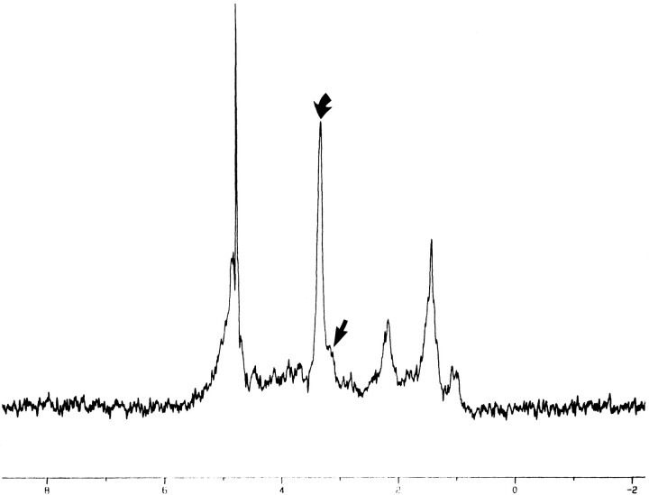
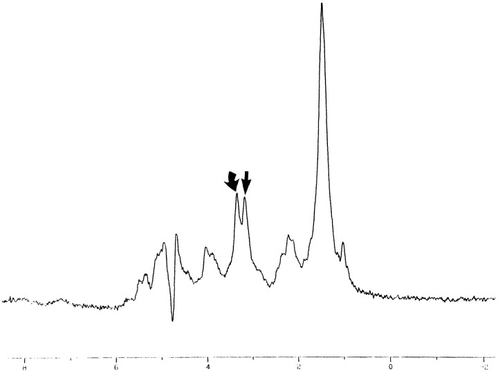
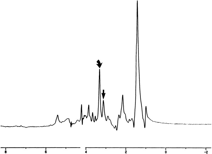
Methods: In vitro one-dimensional proton MR spectroscopy (2,000/136,272 [TR/TE]) was performed at 11 T on tissue specimens obtained from glomus tumors (n = 3), inverting papilloma (n = 1), and schwannoma (n = 1). Cho/Cr area ratios were calculated and compared with similar, previously reported in vitro (11 T) findings and with samples of SCCA and normal muscle.
Results: The Cho/Cr ratio was elevated in relation to muscle in all benign tumors at TE = 136 (glomus tumors = 4.52, inverting papilloma = 3.85, schwannoma = 2.2) and at TE = 272 (glomus tumors = 8.01, inverting papilloma = 2.1, schwannoma = 4.28). The average Cho/Cr ratio for benign lesions was 3.92 (TE = 136) and 6.11 (TE = 272). The Cho/Cr ratio was significantly higher in benign tumors than in both SCCA and muscle. The average Cho/Cr ratio for muscle at TEs of 136 and 272 was 1.16 and 1.31, respectively, whereas for SCCA the average Cho/Cr ratio at TEs of 136 and 272 was 1.67 and 2.45, respectively.
Conclusion: In our small group, the Cho/Cr ratio was significantly higher in benign tumors than in muscle and SCCA of the extracranial head and neck.
Figures


Similar articles
-
In vivo proton MR spectroscopy of primary and nodal nasopharyngeal carcinoma.AJNR Am J Neuroradiol. 2004 Mar;25(3):484-90. AJNR Am J Neuroradiol. 2004. PMID: 15037477 Free PMC article.
-
Proton MR spectroscopy of squamous cell carcinoma of the extracranial head and neck: in vitro and in vivo studies.AJNR Am J Neuroradiol. 1997 Jun-Jul;18(6):1057-72. AJNR Am J Neuroradiol. 1997. PMID: 9194433 Free PMC article.
-
The role of neural networks in improving the accuracy of MR spectroscopy for the diagnosis of head and neck squamous cell carcinoma.AJNR Am J Neuroradiol. 2000 Jun-Jul;21(6):1133-8. AJNR Am J Neuroradiol. 2000. PMID: 10871028 Free PMC article.
-
Proton MR spectroscopy of squamous cell carcinoma of the upper aerodigestive tract: in vitro characteristics.AJNR Am J Neuroradiol. 1996 Sep;17(8):1485-90. AJNR Am J Neuroradiol. 1996. PMID: 8883645 Free PMC article.
-
Preliminary study of 3T 1H MR spectroscopy in bone and soft tissue tumors.Chin Med J (Engl). 2009 Jan 5;122(1):39-43. Chin Med J (Engl). 2009. PMID: 19187615
Cited by
-
In vivo proton MR spectroscopy of primary tumours, nodal and recurrent disease of the extracranial head and neck.Eur Radiol. 2007 Jan;17(1):251-7. doi: 10.1007/s00330-006-0294-2. Epub 2006 May 16. Eur Radiol. 2007. PMID: 16703309
-
In vivo proton MR spectroscopy of primary and nodal nasopharyngeal carcinoma.AJNR Am J Neuroradiol. 2004 Mar;25(3):484-90. AJNR Am J Neuroradiol. 2004. PMID: 15037477 Free PMC article.
-
Imaging Findings of an Epidermoid Cyst Undergoing Malignant Transformation.J Belg Soc Radiol. 2015 Sep 15;99(1):42-45. doi: 10.5334/jbr-btr.829. J Belg Soc Radiol. 2015. PMID: 30039065 Free PMC article.
-
Metabolomics of Head and Neck Cancer: A Mini-Review.Front Physiol. 2016 Nov 8;7:526. doi: 10.3389/fphys.2016.00526. eCollection 2016. Front Physiol. 2016. PMID: 27877135 Free PMC article. Review.
-
Capillary electrophoresis mass spectrometry-based saliva metabolomics identified oral, breast and pancreatic cancer-specific profiles.Metabolomics. 2010 Mar;6(1):78-95. doi: 10.1007/s11306-009-0178-y. Epub 2009 Sep 10. Metabolomics. 2010. PMID: 20300169 Free PMC article.
References
-
- Mafee MF, Barany M, Gotsis ED, et al. Potential use of in vivo proton spectroscopy for head and neck lesions. Radiol Clin N Am 1989;27:243-254 - PubMed
-
- Gill SG, Thomas DGT, Van Bruggen NV, et al. Proton MR spectroscopy of intracranial tumours: in vivo and in vitro studies. J Comput Assist Tomogr 1990;14:497-504 - PubMed
-
- Delikatny EJ, Russell P, Hunter JC, et al. Proton MR and human cervical neoplasia: ex vivo spectroscopy allows distinction of invasive carcinoma of the cervix from carcinoma in situ and other preinvasive lesions. Radiology 1993;188:791-796 - PubMed
Publication types
MeSH terms
Substances
LinkOut - more resources
Full Text Sources
Research Materials