. 2000 Nov-Dec;21(10):1955-61.

Assessment of the deep gray nuclei in holoprosencephaly

Affiliations

Assessment of the deep gray nuclei in holoprosencephaly

E M Simon et al. AJNR Am J Neuroradiol. 2000 Nov-Dec.

Abstract

Background and purpose: Although holoprosencephaly has been known for many years, few detailed analyses have been performed in a large series of patients to outline the range of morphology in this disorder, particularly regarding the deep gray nuclear structures. We reviewed a large patient cohort to elucidate the combinations of morphologic aberrations of the deep gray nuclei and to correlate those findings with recent discoveries in embryology and developmental neurogenetics.

Methods: A retrospective review of the imaging records of 57 patients (43 MR studies and 14 high-quality CT studies) to categorize the spectrum of deep gray nuclear malformations. The hypothalami, caudate nuclei, lentiform nuclei, thalami, and mesencephalon were graded as to their degree of noncleavage. Spatial orientation was also evaluated, as was the relationship of the basal ganglia to the diencephalic structures and mesencephalon. The extent of noncleavage of the various nuclei was then assessed for statistical association.

Results: In every study on which it could be accurately assessed, we found some degree of hypothalamic noncleavage. Noncleavage was also common in the caudate nuclei (96%), lentiform nuclei (85%), and thalami (67%). Complete and partial noncleavage were more common in the caudate nuclei than in the lentiform nuclei. The degree of thalamic noncleavage was uniformly less than that in the caudate and lentiform nuclei. Abnormalities in alignment of the long axis of the thalamus were seen in 71% of cases, and were associated with degree of thalamic noncleavage; 27% of patients had some degree of mesencephalic noncleavage.

Conclusion: The hypothalamus and caudate nuclei are the most severely affected structures in holoprosencephaly, and the mesencephalic structures are more commonly involved than previously thought in this "prosencephalic disorder." These findings suggest the lack of induction of the most rostral aspects of the embryonic floor plate as the cause of this disorder.


Figures
<sc>fig</sc> 1.
fig 1.
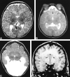
Range of noncleavage of the basal ganglia and thalamus. A, Grade 0: Axial T2-weighted image at the level of the third ventricle shows the abnormally deep anterior IHF (long arrow) and widely separated caudate and lentiform nuclei (short arrows). B, Grade 1: Axial T2-weighted image at the level of the third ventricle shows the medial location and small amount of contact between the caudate nuclei (arrow). Note also the noncleavage of the claustra (arrowheads) and probable anterior limbs of the internal capsules (white matter posterior to claustra). C, Grade 2: Axial T2-weighted image in a more severely affected patient shows the partial noncleavage of the lentiform nuclei (white arrows) and thalami (black arrows). D, Grade 3: Coronal 3D spoiled gradient-echo image through the caudate heads shows complete failure of cleavage (arrows).
<sc>fig</sc> 2.
fig 2.
Range of noncleavage of the hypothalamus. A, Grade 1: Coronal T2-weighted image shows the attenuated anterior recess of the third ventricle (arrow) in the setting of partial hypothalamic noncleavage. B, Grade 2: Coronal T1-weighted image shows the continuity of the hypothalamus across the midline of the basal forebrain (short arrow). Note the associated ectopic neurohypophysis (long arrow). fig 3. Deep gray nuclear mass. Axial T1-weighted image shows the noncleaved deep gray nuclear mass, which is diminished in volume and without discernible structures (arrows)
<sc>fig</sc> 4.
fig 4.
Spatial orientation of the thalami. A, Grade 0: Axial T2-weighted image shows the thalami with their long axes at an angle approximately 30° to 45° relative to the plane of the IHF (arrows). B, Grade 1: Axial T2-weighted image shows the thalami with their long axes approximately parallel to the plane of the IHF (arrows). C, Grade 2: Axial T2-weighted image shows the thalami with their long axes approximately perpendicular to the plane of the IHF (arrows).
<sc>fig</sc> 5.
fig 5.
Diagrams of the developing prosencephalon. A, Superior view of the neural plate, with the top being the rostral end at the anterior neuropore and the bottom being more caudal at the level of the diencephalon. Adapted from with permission from the Annual Review of Neuroscience, Volume 21, 1998. B, Lateral view of the neural plate, with the floor plate (fp) at the bottom and the roof plate (rp) at the top. Adapted from with permission from The Company of Biologists, Ltd. C, Coronal view of the neural plate with the roof plate (rp) (dorsal) at the top and the floor plate (fp) and prechordal plate (pcp) (ventral) at the bottom. The more rostral and alar portion, superiorly (above the horizontal dashed black line in A) and to the left (anterior to the vertical dashed black line in B) is the secondary prosencephalon (sp) whereas the more inferior (in A) and right-sided portion (in B) is the diencephalon (di). The longitudinal gray area represents the prechordal plate. The horseshoe-shaped dashed black line surrounding the prechordal plate (in A) represents the boundary between the basal (or ventral) plate (ba) and the alar (or dorsal) plate (al). Within the secondary prosencephalon, structures at all dorsal and ventral levels are affected. The basal midline regions closest to the floor plate and to the lateral ventricles are the most severely affected in HPE. The reason for this is illustrated in C. In normal embryonic development, an interplay of dorsalizing molecules (emanating from the roof plate) and ventralizing molecules (emanating from the prechordal plate and floor plate) modulates regional identity of tissues along the dorsoventral axis of the neural tube. Either a lack of production of ventralizing factors or an overproduction of dorsalizing factors can result in noncleavage (commonly called fusion) of structures that normally lie just lateral to the midline. This is the presumed mechanism by which HPE develops. nh indicates neurohypophysis; hy, hypothalamus; ey, eyefields; gp, globus pallidus; am, amygdala; ac, anterior commissure; lt, laminal terminalis; st, striatum (caudate and putamen); cx, cortex; hi, hippocampus; se, septum; po, preoptic nucleus; ob, olfactory bulb; bst, bed nucleus of stria terminalis.

References

    1. DeMyer W, Zeman W. Alobar holoprosencephaly (arhinencephaly) with median cleft lip and palate: clinical, nosologic, and electroencephalographic considerations. Confin Neurol 1963;23:1-36 - PubMed
    1. DeMyer W, Zeman W, Palmer CG. The face predicts the brain: diagnostic significance of median facial anomalies for holoprosencephaly (arhinencephaly). Pediatrics 1964;34:256-263 - PubMed
    1. Probst FP. The Prosencephalies: Morphology, Neuroradiological Appearances and Differential Diagnosis.. New York: Springer; 1979
    1. Kobori JA, Herrick MK, Urich H. Arhinencephaly: the spectrum of associated malformations. Brain 1987;110:237-260 - PubMed
    1. Sulik KK, Johnston MC. Embryonic origin of holoprosencephaly: interrelationship of the developing brain and face. Scan Elec Microsc 1982;260:309-322 - PubMed

Publication types

LinkOut - more resources