. 2001 Mar 23;276(12):9366-74.
doi: 10.1074/jbc.M009897200. Epub 2000 Dec 8.

A role for poly(ADP-ribose) polymerase in the transcriptional regulation of the melanoma growth stimulatory activity (CXCL1) gene expression

Affiliations

A role for poly(ADP-ribose) polymerase in the transcriptional regulation of the melanoma growth stimulatory activity (CXCL1) gene expression

C Nirodi et al. J Biol Chem. .

Abstract

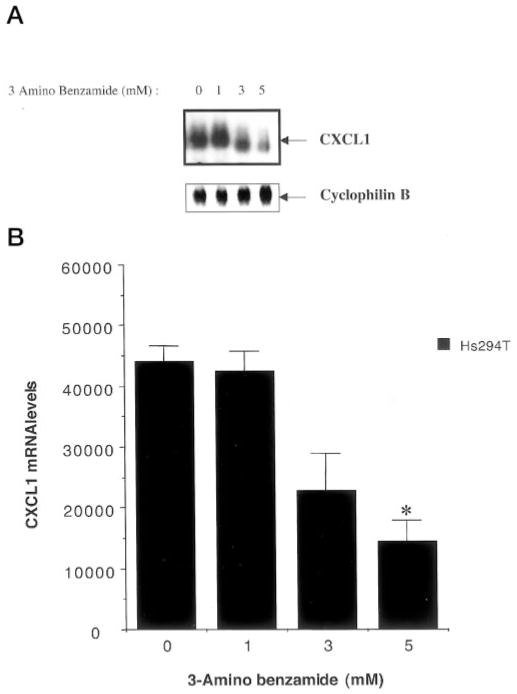
The melanoma growth stimulatory activity/growth-regulated protein, CXCL1, is constitutively expressed at high levels during inflammation and progression of melanocytes into malignant melanoma. It has been shown previously that CXCL1 overexpression in melanoma cells is due to increased transcription as well as stability of the CXCL1 message. The transcription of CXCL1 is regulated through several cis-acting elements including Sp1, NF-kappaB, HMGI(Y), and the immediate upstream region (IUR) element (nucleotides -94 to -78), which lies immediately upstream to the nuclear factor kappaB (NF-kappaB) element. Previously, it has been shown that the IUR is necessary for basal and cytokine-induced transcription of the CXCL1 gene. UV cross-linking and Southwestern blot analyses indicate that the IUR oligonucleotide probe selectively binds a 115-kDa protein. In this study, the IUR element has been further characterized. We show here that proximity of the IUR element to the adjacent NF-kappaB element is critical to its function as a positive regulatory element. Using binding site oligonucleotide affinity chromatography, we have selectively purified the 115-kDa IUR-F. Mass spectrometry/mass spectrometry/matrix-assisted laser desorption ionization/time of flight spectroscopy and amino acid analysis as well as microcapillary reverse phase chromatography electrospray ionization tandem mass spectrometry identified this protein as the 114-kDa poly(ADP-ribose) polymerase (PARP1). Furthermore, 3-aminobenzamide, an inhibitor of PARP-specific ADP-ribosylation, inhibits CXCL1 promoter activity and reduces levels of CXCL1 mRNA. The data point to the possibility that PARP may be a coactivator of CXCL1 transcription.


Figures

Fig. 1
Fig. 1. The IUR element is a positive cis element, which requires the TCGAT motif as well as contiguity with the adjacent NF-κB element
A, 5 × 10⁵ Hs294T cells were transfected with RSV-β-gal vector and one of the following luciferase reporter constructs: 1, a reporter construct lacking the CXCL1 promoter (pGL2); 2, a reporter construct driven by the wild-type CXCL1 promoter in the correct orientation relative to the transcription start site (WT); 3, a reporter construct with the CXCL1 promoter in the opposite direction (REV); 4, a mutant CXCL1 promoter in which the TCGAT motif was altered to AGTAC (mIUR). B, 5 × 10⁵ Hs294T cells were transfected with RSV-β-gal vector as well as luciferase reporter constructs driven by the wild type CXCL1 promoter or mutant promoter which had an insertion of 2 bp (IN:2), 6 bp (IN:6), 12 bp (IN:12), or 25 bp (IN:25) between the IUR and the NF-κB elements. 48 h after transfection, cells were harvested and luciferase activity was measured and normalized to β-galactosidase activity. Shown here is a representative of three independent experiments performed each time in triplicate. Mean normalized values obtained from the sample showing the highest inhibition in promoter activity were compared with those from samples transfected with the wild type promoter and were found to be significantly different according to Student’s t test (paired). The asterisk (*) indicates p < 0.01.
Fig. 2
Fig. 2. Binding site oligonucleotide affinity purification of the 115-kDa IUR-specific protein
In electrophoretic mobility shift assays (A), crude HeLa nuclear extract (2 μg) (lane 1) or 200 ng of bound fractions eluting at 0.2 M NaCl (lanes 2–5), 0.4 M NaCl (lanes 7–10), 0.6 M NaCl (lanes 11–14), or 1 M NaCl (lanes 16–18) from the IUR-agarose column, were tested for binding to the 2R probe in EMSA. All samples contained a 250-fold molar excess of cold oligonucleotide corresponding to the 2mR probe. B, Southwestern blot analysis: Polypeptides in crude HeLa nuclear extract (25 μg) (lanes 1 and 3) or the 0.4 M NaCl fraction (100 ng) eluted from the IUR-agarose column were separated on 8% SDS-PAGE gels, trans-blotted on nitrocellulose membranes, and probed with radiolabeled oligonucleotides corresponding to either the 2R (lanes 1 and 2) or the 2mR (lanes 3 and 4) probes. The relative molecular size is indicated on the left. C, silver-stained SDS-PAGE profile of the 0.4 M NaCl fraction. Analysis: polypeptides in crude HeLa nuclear extract (500 ng) or the 0.4 M NaCl fraction (100 ng) eluted from the IUR-agarose column was separated on 8% SDS-PAGE gels and the protein was visualized by silver-staining. D, SDS-PAGE electro-elution. The 0.4 M IUR-agarose eluate was then fractionated by electro-elution through a 7% SDS-PAGE using the model 491 Prep Cell (Bio-Rad). Fractions were collected, every eighth fraction was electrophoresed on an 8% SDS-PAGE gel, and proteins were stained by silver staining.
Fig. 3
Fig. 3. PARP can bind the IUR element
A, Western blot analysis. Crude HeLa nuclear extract (25 μg), unbound/flow-through fraction from the IUR-agarose column (25 μg) and 100 ng of 0.2 M (lanes 3 and 4) or 0.4 M (lanes 5 and 6) IUR-agarose fractions were separated on 8% SDS-PAGE gels and probed with anti-PARP antibody. Arrow (→) indicates the relative mobility of the PARP protein. B, EMSA. Partially purified, commercially available PARP was tested for binding to the 2xIUR (lanes 1–3) or the 2xmIUR (lanes 4–6) probes. Nonspecific complexes were competed with poly(dI-dC) in the range of 0.2- 1.0 μg. The arrow (→) indicates the specific complex formed by PARP with the 2xIUR probe, and NS represents nonspecific complexes.
Fig. 4
Fig. 4. 3-AB inhibits MGSA/GRO expression
A, Northern analysis. Hs294T cells were treated for 48 h with indicated concentrations of 3-AB. RNA from samples was resolved on a 1.4% formaldehydeagarose gel, transblotted to nitrocellulose membrane, and probed with an MGSA/GRO cDNA probe (A, top panel). Blots were stripped and reprobed with a cDNA probe for cyclophilin B transcripts (A, bottom panel). Blots were densitometrically scanned using a PhosphorImager (Molecular Dynamics). Values for MGSA/GRO mRNA were normalized against those for cyclophilin mRNA. B, a graphical representative of six independent experiments, each performed in triplicate is shown. The results in all six experiments were qualitatively identical. Error bars represent S.D. values. The mean normalized value obtained from samples receiving the highest dose of 3-AB was compared with those from samples receiving Me2SO and were found to be significantly different according to Student’s paired t test. The asterisk indicates p < 0.01.
Fig. 5
Fig. 5. 3-AB inhibits CXCL1 promoter activity
5 × 10⁵ cells were first transfected with 1 μg of pRSV-β-gal reporter as well as reporter constructs driven by either the wild type (MGSA.Luc) or the mutant CXCL1 promoter (mIUR.Luc). Six hours after transfection, the medium was replaced by medium containing either Me2SO or 3 -AB within a concentration range of 1–5 mM. Cells were harvested 48 h after transfection and luciferase activity was measured. Values obtained were normalized to β-galactosidase activity. The experiment was performed three times in triplicate. Error bars represent standard deviations. The mean normalized values obtained from samples receiving the highest dose of 3-AB as compared with those from samples receiving Me2SO were significantly different according to Student’s paired t test The asterisk indicates p < 0.01.
Fig. 6
Fig. 6. 3-AB has no effect on binding to the IUR element
Hs294T cells were treated with either Me2SO alone or with 1–5 mM 3-AB for 48 h. Cells were harvested, and nuclear extracts were made. 10 μg of nuclear extract was incubated with 32P-labeled 2xIUR oligonucleotide probe in the presence of 1–2 μg of poly(dI-dC). The reaction mixtures electrophoresed on 6% native polyacrylamide gels, which were then dried and processed for autoradiography. The arrow indicates the specific complex associated with the IUR probe. Shown here is a representative of three independent experiments. Results were qualitatively similar in all three experiments.

Similar articles

Cited by

References

    1. Richmond A, Shattuck RL. In: Chemoattractant Ligands and Their Receptors. Horuk R, editor. CRC Press; Boca Raton, FL: 1996. pp. 87–124.
    1. Shattuck RL, Wood LD, Jaffe GJ, Richmond A. Mol Cell Biol. 1994;14:791–802. - PMC - PubMed
    1. Wood LD, Farmer AA, Richmond A. Nucleic Acids Res. 1995;23:4210–4219. - PMC - PubMed
    1. Wang XC, Jobin C, Allen JB, Roberts WL, Jaffe GJ. Invest Ophthal Vis Sci. 1999;40:477–486. - PubMed
    1. Shattuck-Brandt RL, Richmond A. Cancer Res. 1997;57:3032–3039. - PubMed

Publication types

MeSH terms

LinkOut - more resources