. 2001 Jan;21(1):224-34.
doi: 10.1128/MCB.21.1.224-234.2001.

Defining roles for HOX and MEIS1 genes in induction of acute myeloid leukemia

Affiliations

Defining roles for HOX and MEIS1 genes in induction of acute myeloid leukemia

U Thorsteinsdottir et al. Mol Cell Biol. 2001 Jan.

Abstract

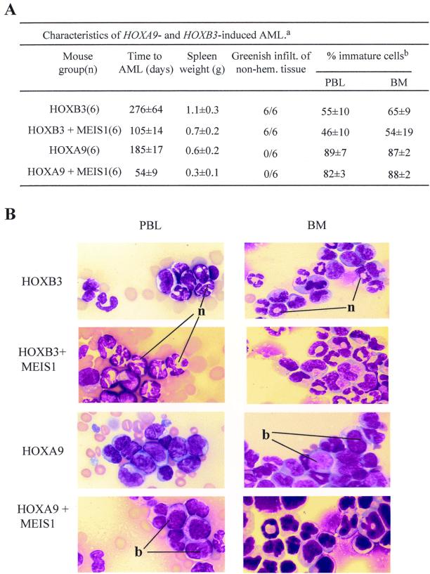
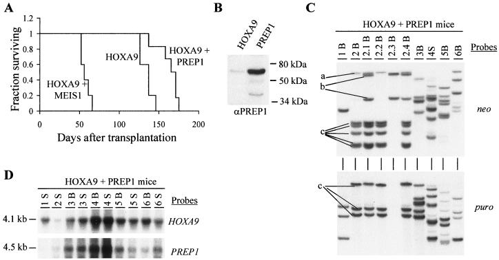
Complex genetic and biochemical interactions between HOX proteins and members of the TALE (i.e., PBX and MEIS) family have been identified in embryonic development, and some of these interactions also appear to be important for leukemic transformation. We have previously shown that HOXA9 collaborates with MEIS1 in the induction of acute myeloid leukemia (AML). In this report, we demonstrate that HOXB3, which is highly divergent from HOXA9, also genetically interacts with MEIS1, but not with PBX1, in generating AML. In addition, we show that the HOXA9 and HOXB3 genes play key roles in establishing all the main characteristics of the leukemias, while MEIS1 functions only to accelerate the onset of the leukemic transformation. Contrasting the reported functional similarities between PREP1 and MEIS1, such as PBX nuclear retention, we also show that PREP1 overexpression is incapable of accelerating the HOXA9-induced AML, suggesting that MEIS1 function in transformation must entail more than PBX nuclear localization. Collectively, these data demonstrate that MEIS1 is a common leukemic collaborator with two structurally and functionally divergent HOX genes and that, in this collaboration, the HOX gene defines the identity of the leukemia.


Figures

FIG. 1
FIG. 1
Diagrammatic representation of the HOXA9, HOXB3, MEIS1a, and PREP1 proteins and the retroviral constructs used in this study. (A) Sequence comparison of the HOXA9 and HOXB3 proteins and the MEIS1a and the PREP1 proteins used in this study. Both HOXA9 and HOXB3 proteins have a motif (ANWL in HOXA9 and YPWM in HOXB3) N-terminal to the homeodomain that is essential for their interaction with PBX proteins. Apart from their homeodomains, which are 70% identical, these proteins do not display significant sequence similarity. The MEIS1a and the PREP1 proteins share sequence similarity only in their homeodomains (70%) and in the N-terminal HMA (60%) and HMB (60%) domains that mediate interactions with PBX proteins. (B) Diagrammatic representation of the integrated MSCV-HOXA9, MSCV-HOXB3, MSCV-PBX1, MSCV-MEIS1, and MSCV-PREP1 proviruses. The expected sizes of the full-length long terminal repeat (LTR)-driven viral transcripts are shown. Restriction sites indicated are KpnI (Kp) (shown only for the HOXA9 virus but present in all constructs) and EcoRI (E). HD, homeodomain; HM, Homothorax-Meis domain.
FIG. 2
FIG. 2
Demonstration of collaboration between HOXB3 and MEIS1, but not HOXB3 and PBX1, in leukemogenesis. (A) Survival graph demonstrating the collaboration between HOXB3 and MEIS1, but not PBX1, in the development of AML. The survival of the HOXB3-MEIS1 mice was significantly shorter than that of the HOXB3 mice (P < 0.001, two-tailed Student's t test) and the HOXB3-PBX1 mice (P < 0.007). The survival of the HOXB3-PBX1 mice was not significantly different from that of the HOXB3 mice. (B) Southern blot analyses of genomic DNA isolated from the bone marrow and/or spleen of the HOXB3-PBX1 and HOXB3-MEIS1 chimeras. DNA was digested with KpnI to release the integrated HOXB3 (4.3-kb), MEIS1 (4.2-kb), or PBX1 (4.5-kb) proviral fragments. The membranes were hybridized with a neo-specific probe to detect the HOXB3 provirus and a puro-specific probe to detect the MEIS1 or PBX1 provirus. (C) Northern blot analysis of total RNA (10 μg) isolated from bone marrow or spleen cells of the HOXB3-PBX1 and HOXB3-MEIS1 mice. The membranes were hybridized with full-length HOXB3, MEIS1, or PBX1 cDNA probes. (D) Southern blot analysis of DNA isolated from bone marrow of primary and secondary HOXB3-MEIS1 mice. The DNA was digested with EcoRI, which cuts the integrated provirus once, thus generating a unique fragment for each proviral integration site. The membranes were hybridized first with a neo-specific probe for detection of the HOXB3 proviral fragment(s) (top panel) and then subsequently with a puro-specific probe to detect the MEIS1 proviral fragment(s) (bottom panel). In panels B, C, and D, each primary recipient is identified with a specific number and its secondary recipients or cell lines generated from each primary recipient, with a derivative thereof (e.g., 1.1 and 1.2 and CL1, CL2, etc.). B, bone marrow; S, spleen; CL, cell lines.
FIG. 3
FIG. 3
Overexpression of MEIS1 is not permissive for B-lymphoid development but neither induces proliferation of bone marrow cells nor predisposes recipients to lymphoid or myeloid leukemias. (A) Survival graph of chimeras reconstituted with HOXA9-, HOXB3-, MEIS1-, or PBX1-transduced bone marrow cells, demonstrating, for the observation period of 450 days, that only the chimeras engineered to overexpress HOXB3 or HOXA9, but not MEIS1 or PBX1, developed leukemia. (B) Southern blot analyses of genomic DNA isolated from the bone marrow and spleen of puro-control, PBX1, and MEIS1 mice. DNA was digested with KpnI to release the integrated puro (2.7-kb), MEIS1 (4.2-kb), or PBX1 (4.5-kb) proviral fragments. The membranes were hybridized with a puro-specific probe to detect the control, MEIS1, and PBX1 proviruses. (C) Flow cytometric analysis of hematopoietic cells from bone marrow, spleen, and thymus of EGFP control and MEIS1-EYFP mice transplanted 60 days earlier with EGFP- or MEIS1-EYFP-transduced bone marrow cells, respectively. Numbers in the inset quadrant represent the percentages of live cells in the corresponding quadrant. (D) In vitro proliferation of HOXA9-EGFP (▴)-, EGFP-control (□)-, and MEIS1-EYFP (●)-positive bone marrow cells isolated from corresponding mouse chimeras at 60 days after transplantation. B, bone marrow; S, spleen; BMT, bone marrow transplantation.
FIG. 4
FIG. 4
Differences between HOXB3- and HOXA9-induced AMLs. (A) Main characteristics of the AMLs that developed in HOXB3, HOXB3-MEIS1, HOXA9, and HOXA9-MEIS1 bone marrow chimeras. a, Results are expressed as the means ± standard deviations for the indicated number of mice. b, Determination of the proportion of immature and mature cells in hematopoietic tissue of the leukemic mice was based on morphological criteria, i.e., mature cells with segmented nuclei and immature cells, blast-like. For each tissue sample, n = 200 cells were counted from n = 3 representative mice in each group. infilt., infiltration; non-hem., nonhematopoietic. (B) Wright staining of peripheral blood smears (PBL) and bone marrow (BM) cytospins from representative leukemic HOXB3, HOXB3-MEIS1, HOXA9, and HOXA9-MEIS1 mice. Magnification, ×100 for all. n, neutrophil; b, blast.
FIG. 5
FIG. 5
Demonstration of lack of collaboration between HOXA9 and PREP1 in leukemogenesis. (A) Survival graph demonstrating that co-overexpression of PREP1 with HOXA9, in contrast to that with MEIS1, does not accelerate the occurrence of the HOXA9-induced AML. The survival of the HOXA9-MEIS1 mice was significantly shorter than that of the HOXA9 mice (P < 0.001, two-tailed Student's t test) and the HOXA9-PREP1 mice (P < 0.001). (B) Western blot analysis of total-cell lysates from the HOXA9 and PREP1 viral producer cells. The membrane was probed with rabbit anti-human PREP1 polyclonal antibody. The position of the full-length 64-kDa PREP1 protein is indicated. Two minor products, as previously described (5), are also detected (one generated by an internal ATG site). (C) Southern blot analysis of DNA isolated from bone marrow of primary and secondary HOXA9-PREP1 mice. The DNA was digested with EcoRI, which cuts the integrated provirus once, thus generating a unique fragment for each proviral integration site. The membranes were hybridized first with a neo-specific probe for the detection of the HOXA9 proviral fragment(s) (top panel) and subsequently with a puro-specific probe to detect the PREP1 proviral fragment(s) (bottom panel). For clarity, the three different clones detected in the primary and secondary recipients of mouse 2 are labeled a, b, and c. (D) Northern blot analysis of total RNA (10 μg) isolated from bone marrow and spleen cells of the HOXA9-PREP1 mice. The membranes were hybridized with full-length HOXA9 and PREP1 cDNA probes. In panels C and D, each primary recipient is identified with a specific number, and its secondary recipients are identified with a derivative thereof (e.g., 1.1, 1.2, etc.). B, bone marrow; S, spleen.

Similar articles

Cited by

References

    1. Abu-Shaar M, Ryoo H D, Mann R S. Control of the nuclear localization of extradenticle by competing nuclear import and export signals. Genes Dev. 1999;13:935–945. - PMC - PubMed
    1. Azpiazu N, Morata G. Functional and regulatory interactions between Hox and extradenticle genes. Genes Dev. 1998;12:261–273. - PMC - PubMed
    1. Berthelsen J, Kilstrup-Nielsen C, Blasi F, Mavilio F, Zappavigna V. The subcellular localization of PBX1 and EXD proteins depends on nuclear import and export signals and is modulated by association with PREP1 and HTH. Genes Dev. 1999;13:946–953. - PMC - PubMed
    1. Berthelsen J, Zappavigna V, Feretti E, Mavilio F, Blasi F. The novel homeoprotein Prep1 modulates Pbx-Hox protein cooperativity. EMBO J. 1998;17:1434–1445. - PMC - PubMed
    1. Berthelsen J, Zappavigna V, Mavilio F, Blasi F. Prep1, a novel functional partner of Pbx proteins. EMBO J. 1998;17:1423–1433. - PMC - PubMed

Publication types

MeSH terms

LinkOut - more resources