. 2001 Jan 2;98(1):271-6.
doi: 10.1073/pnas.98.1.271.

Malaria parasite exit from the host erythrocyte: a two-step process requiring extraerythrocytic proteolysis

Affiliations

Malaria parasite exit from the host erythrocyte: a two-step process requiring extraerythrocytic proteolysis

B L Salmon et al. Proc Natl Acad Sci U S A. .

Abstract

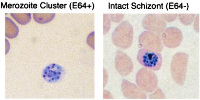
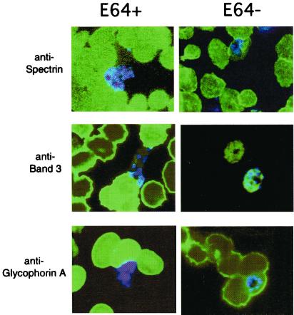
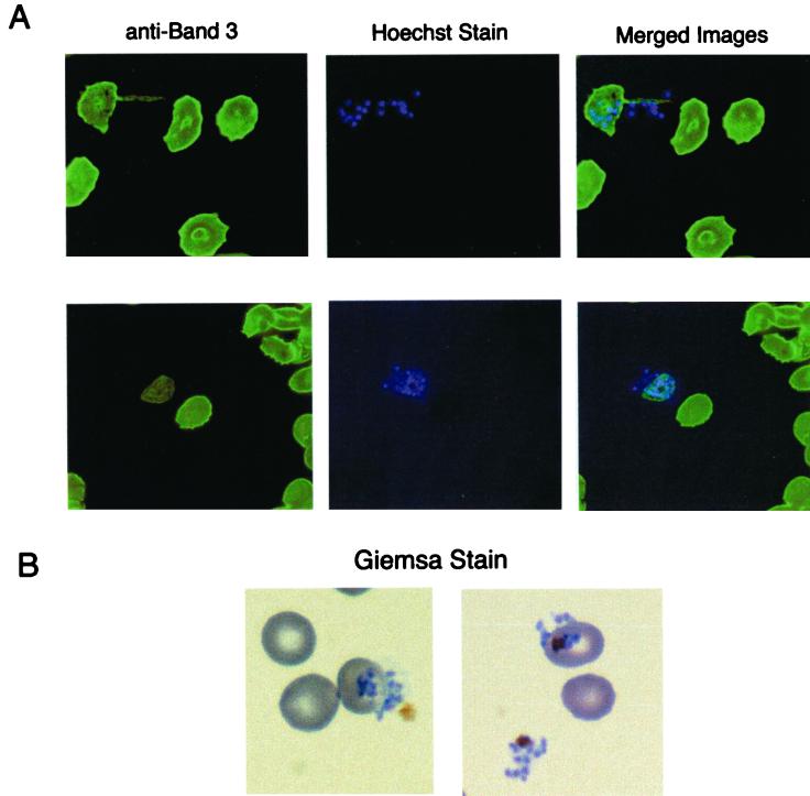
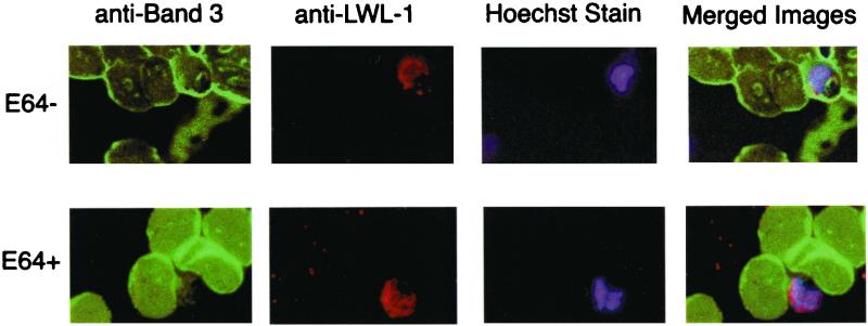
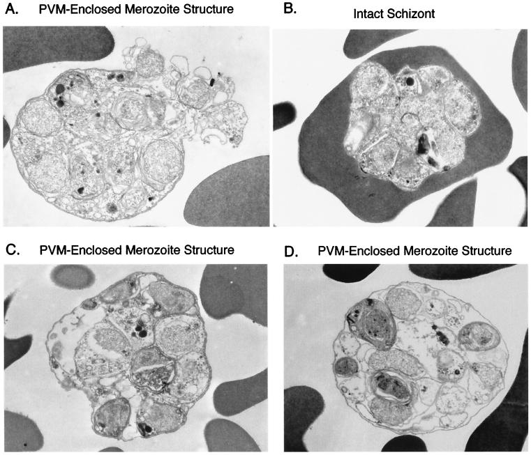
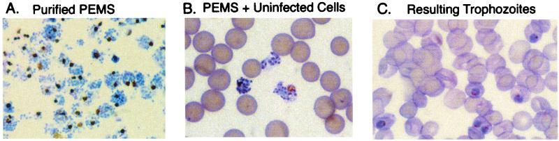
Intraerythrocytic malaria parasites replicate by the process of schizogeny, during which time they copy their genetic material and package it into infective merozoites. These merozoites must then exit the host cell to invade new erythrocytes. To better characterize the events of merozoite escape, erythrocytes containing Plasmodium falciparum schizonts were cultured in the presence of the cysteine protease inhibitor, l-transepoxy-succinyl-leucylamido-(4-guanidino)butane (E64). This treatment resulted in the accumulation of extraerythrocytic merozoites locked within a thin, transparent membrane. Immunomicroscopy demonstrated that the single membrane surrounding the merozoites is not erythrocytic but rather is derived from the parasitophorous vacuolar membrane (PVM). Importantly, structures identical in appearance can be detected in untreated cultures at low frequency. Further studies revealed that (i) merozoites from the PVM-enclosed merozoite structures (PEMS) are invasive, viable, and capable of normal development; (ii) PEMS can be purified easily and efficiently; and (iii) when PEMS are added to uninfected red blood cells, released merozoites can establish a synchronous wave of infection. These observations suggest that l-transepoxy-succinyl-leucylamido-(4-guanidino)butane (E64) causes an accumulation of an intermediate normally present during the process of rupture. We propose a model for the process of rupture: merozoites enclosed within the PVM first exit from the host erythrocyte and then rapidly escape from the PVM by a proteolysis-dependent mechanism.


Figures

Figure 1
Figure 1
Effect of E64 treatment on intraerythrocytic schizonts. Synchronous cultures containing middle-stage schizonts were cultured in the presence of 10 μM E64 for approximately 8 h (Left). Results were visualized by light microscopy of Giemsa-stained blood smears. An untreated culture of late-stage schizonts is provided for comparison (Right).
Figure 2
Figure 2
Localization of erythrocytic proteins and merozoites in schizont-containing blood smears cultured in the presence or absence of E64. Synchronous cultures of middle-stage schizonts were cultured in the presence of 10 μM E64 for approximately 8 h (E64+) or served as untreated controls (E64−). Blood smears of resulting cultures were reacted with monoclonal antibodies directed against spectrin (IgG diluted 1:500), band 3 (IgG diluted 1:5,000), and glycophorin A (IgM diluted 1:800) and then reacted with secondary antibodies FITC-conjugated goat anti-mouse IgG or IgM. Nuclear staining of merozoites was detected by Hoechst stain. Resulting images were visualized by using a Zeiss microscope and merged to compare nuclear stain versus antibody localization patterns.
Figure 3
Figure 3
Analysis of E64-treated parasites in the process of exiting the host cell. (A) Blood smears were reacted with antibody directed against band 3 (1:5,000) and stained with Hoescht stain to detect nuclei of merozoites. Resulting images were visualized by using a Zeiss microscope and merged to compare localization patterns. (B) In parallel, blood smears were Giemsa-stained and viewed by light microscopy.
Figure 4
Figure 4
PVM staining is extraerythrocytic in parasites treated with E64. Erythrocytes containing synchronous cultures of middle-stage schizonts were cultured in the presence of E64 for 8 h (E64+) or served as untreated controls (E64−). Blood smears were reacted with primary antibodies against the erythrocyte membrane marker band 3 (IgG 1:5,000) and the PVM marker LWL-1 (IgM 1:5) and then reacted with secondary antibodies FITC-conjugated goat anti-mouse IgG and rhodamine-conjugated goat anti-mouse IgM. Nuclei of merozoites were detected by using Hoechst stain. Resulting images were viewed by using a Zeiss microscope and merged to compare localization patterns.
Figure 5
Figure 5
Electron microscopy analysis of the effect of E64 on schizont maturation. Synchronous cultures of middle-stage schizonts were cultured in the presence of 10 μM E64 for approximately 8 h (A, C, and D), or late-stage schizonts were harvested to serve as a control (B). Micrographs were viewed by using a Zeiss 902 electron microscope.
Figure 6
Figure 6
Giemsa-stained smears of purified PEMS, PEMS added to uninfected erythrocytes, and resulting trophozoites. PEMS were allowed to accumulate in E64-treated cultures of schizonts. Resulting PEMS were purified on a Percoll cushion and added to uninfected erythrocytes. Blood smears 36 h postinfection were stained with Giemsa and viewed by light microscopy.

Comment in

References

    1. Trigg P I, Kondrachine A V. In: Malaria: Parasite Biology, Pathogenesis and Protection. Sherman I W, editor. Washington, DC: Am. Soc. Microbiol.; 1998. pp. 11–22.
    1. Lyon A J, Haynes J D. J Immunol. 1986;136:2245–2251. - PubMed
    1. Rosenthal P J, Kim K, McKerrow J H, Leech J H. J Exp Med. 1987;166:816–821. - PMC - PubMed
    1. Delplace P, Dubremetz J F, Fortier B, Vernes A. Mol Biochem Parasitol. 1985;17:239–251. - PubMed
    1. Bzik D J, Li W B, Horii T, Inselburg J. Mol Biochem Parasitol. 1988;30:279–288. - PubMed

Publication types

MeSH terms

LinkOut - more resources