. 2000 Dec 19;97(26):14680-5.
doi: 10.1073/pnas.011510698.

A recombinant rabies virus expressing vesicular stomatitis virus glycoprotein fails to protect against rabies virus infection

Affiliations

A recombinant rabies virus expressing vesicular stomatitis virus glycoprotein fails to protect against rabies virus infection

H D Foley et al. Proc Natl Acad Sci U S A. .

Abstract

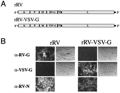
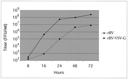
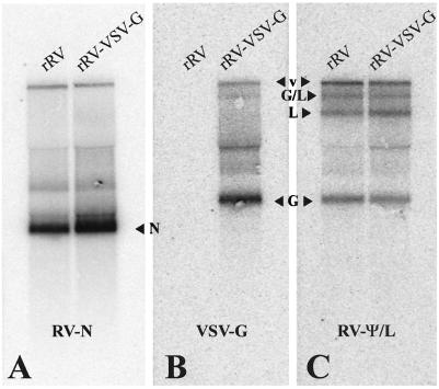
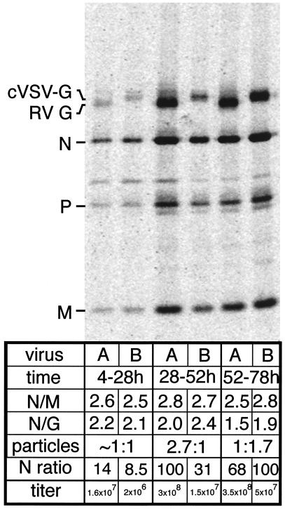
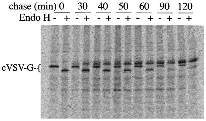
To investigate the importance of the rabies virus (RV) glycoprotein (G) in protection against rabies, we constructed a recombinant RV (rRV) in which the RV G ecto- and transmembrane domains were replaced with the corresponding regions of vesicular stomatitis virus (VSV) glycoprotein (rRV-VSV-G). We were able to recover rRV-VSV-G and found that particle production was equal to rRV. However, the budding of the chimeric virus was delayed and infectious titers were reduced 10-fold compared with the parental rRV strain containing RV G. Biochemical analysis showed equal replication rates of both viruses, and similar amounts of wild-type and chimeric G were present in the respective viral particles. Additional studies were performed to determine whether the immune response against rRV-VSV-G was sufficient to protect against rabies. Mice were primed with rRV or rRV-VSV-G and challenged with a pathogenic strain of RV 12 days later. Similar immune responses against the internal viral proteins of both viruses indicated successful infection. All mice receiving the rRV vaccine survived the challenge, whereas immunization with rRV-VSV-G did not induce protection. The results confirm the crucial role of RV G in an RV vaccine.


Figures

Figure 1
Figure 1
(A) Genome of rRV and rRV-VSV-G. RV SAD L16 was modified to contain a SmaI site upstream of the RV G gene and a NheI site upstream of the ψ region. rRV-VSV-G was created by replacing the RV G ecto- and transmembrane domains with those of VSV G by using a newly created HpaI site. (B) Expression of RV G and VSV G by rRVs. BSR cells were infected with rRV or rRV-VSV-G (moi of 0.1) and immunostained 48 h later with anti-RV-G, anti-VSV-G, or anti-RV-N antibody. Light field pictures are shown for anti-RV-G and anti-VSV-G.
Figure 2
Figure 2
One-step growth curves of rRV and rRV-VSV-G. BSR cells were infected with an moi of 10 with either rRV (♦) or rRV-VSV-G (▪). Supernatant (100 μl) was taken at 8, 16, 24, 48, and 72 h after infection and titered in duplicate.
Figure 3
Figure 3
Northern blot analysis of rRVs. BSR cells were infected with rRV or rRV-VSV-G (moi of 1), and total RNA was isolated 24 h later. RNA was separated by electrophoresis and transferred to a nylon membrane that was hybridized with a probe specific for RV-N (A), VSV-G (B), or a cDNA fragment spanning the entire RV-ψ and a portion of the RV-L gene (ψ + L) (C). G/L = RV G + L bicistronic RNA, L = RV L, G = glycoprotein, N = RV-N.
Figure 4
Figure 4
Comparison of foci size between rRV and rRV-VSV-G. BSR cells were infected with rRV and rRV-VSV-G (moi of 0.1). Cells were fixed 48 h after infection and immunostained with an RV-N-specific antibody. (A) rRV. (B) rRV-VSV-G.
Figure 5
Figure 5
Particle production and infectious titers of rRV and rRV-VSV-G. BSR cells were infected with rRV (A) or rRV-VSV-G (B) at an moi of 50 and labeled with [35S]methionine for 24 h over 4–28, 28–52, or 52–78 h. Labeled virions were purified twice over 20% (vol/vol) sucrose and disrupted. Then the viral proteins were separated by SDS/PAGE. Gels were analyzed by PhosphorImaging, and viral proteins were quantified. The ratio of N protein to M protein (N/M) per virus was determined along with the ratios of N protein to G protein (N/G). The lane with the highest amount of N protein was determined for each virus and set to equal 100 (N ratio lane) and the other values were calculated in proportion to 100. The N ratios of rRV and rRV-VSV-G were compared and estimated in the particles row. A portion of the initial supernatant was used to determine the infectious titer at each time point (titer).
Figure 6
Figure 6
Transport kinetics of cVSV-G. Infected cells were pulsed with [35S]methionine for 10 min and chased for 0, 30, 40, 50, 60, 90, or 120 min. Cell extracts were incubated with or without endo H for 4 h at 37°C and immunoprecipitated with an RV G endoplasmic tail-specific antibody. Proteins were separated by SDS/PAGE and analyzed by PhosphorImaging. Endo H + or − indicates with or without endo H.
Figure 7
Figure 7
ELISA analysis of mouse sera against RV RNP and RV G. Sera were collected 9 days after priming from all mice and analyzed for reactivity against RV G (A) or RV RNP (B). Sera from each mouse were run in duplicate. Graphs represent the average of 10 mice per group with the error bars representing standard deviations.

References

    1. Cox J H, Dietzschold B, Schneider L G. Infect Immun. 1977;16:754–759. - PMC - PubMed
    1. Wiktor T J, Gyorgy E, Schlumberger D, Sokol F, Koprowski H. J Immunol. 1973;110:269–276. - PubMed
    1. Perrin P, Thibodeau L, Sureau P. Vaccine. 1985;3:325–332. - PubMed
    1. Macfarlan R I, Dietzschold B, Koprowski H. Mol Immunol. 1986;23:733–741. - PubMed
    1. Celis E, Ou D, Dietzschold B, Koprowski H. J Virol. 1988;62:3128–3134. - PMC - PubMed

Publication types

MeSH terms

LinkOut - more resources