. 2000 Dec 19;97(26):14614-9.
doi: 10.1073/pnas.250289597.

Autosomal dominant myopathy: missense mutation (Glu-706 --> Lys) in the myosin heavy chain IIa gene

Affiliations

Autosomal dominant myopathy: missense mutation (Glu-706 --> Lys) in the myosin heavy chain IIa gene

T Martinsson et al. Proc Natl Acad Sci U S A. .

Abstract

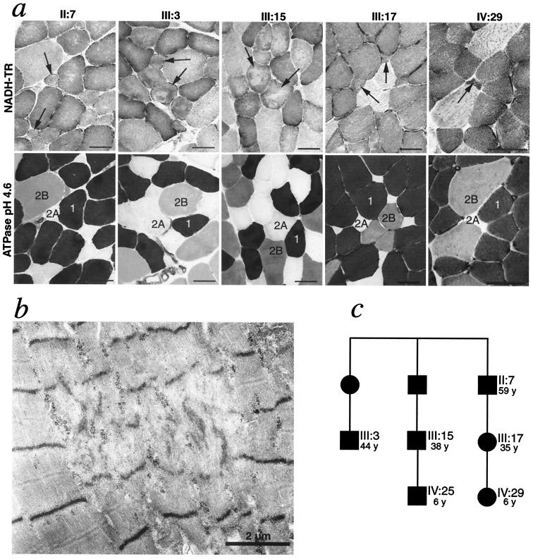
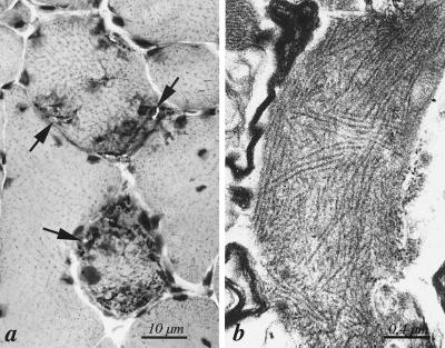
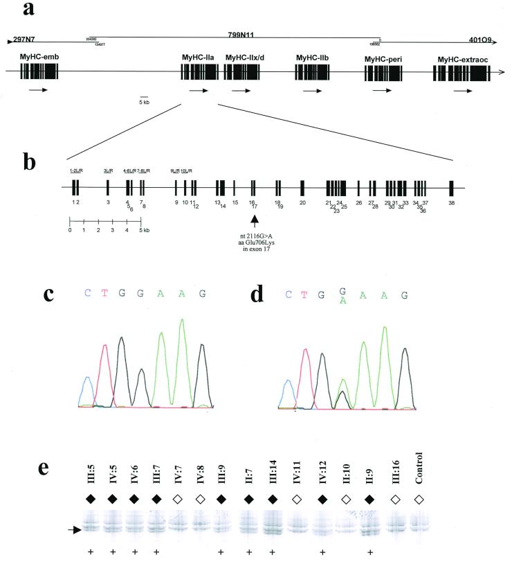
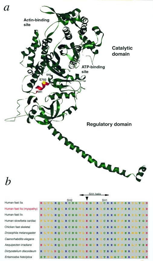
We here report on a human myopathy associated with a mutation in a fast myosin heavy chain (MyHC) gene, and also the genetic defect in a hereditary inclusion body myopathy. The disorder has previously been described in a family with an "autosomal dominant myopathy, with joint contractures, ophthalmoplegia, and rimmed vacuoles." Linkage analysis and radiation hybrid mapping showed that the gene locus (Human Genome Map locus name: IBM3) is situated in a 2-Mb region of chromosome 17p13, where also a cluster of MyHC genes is located. These include the genes encoding embryonic, IIa, IIx/d, IIb, perinatal, and extraocular MyHCs. Morphological analysis of muscle biopsies from patients from the family indicated to us that the type 2A fibers frequently were abnormal, whereas other fiber types appeared normal. This observation prompted us to investigate the MyHC-IIa gene, since MyHC-IIa is the major isoform in type 2A fibers. The complete genomic sequence for this gene was deduced by using an "in silico" strategy. The gene, found to consist of 38 exons, was subjected to a complete mutation scan in patients and controls. We identified a missense mutation, Glu-706 --> Lys, which is located in a highly conserved region of the motor domain, the so-called SH1 helix region. By conformational changes this region communicates activity at the nucleotide-binding site to the neck region, resulting in the lever arm swing. The mutation in this region is likely to result in a dysfunctional myosin, compatible with the disorder in the family.


Figures

Figure 1
Figure 1
Analysis of involvement of different muscle fiber types. (a) Analysis by enzyme histochemical staining of myofibrillar ATPase (Lower) and NADH-tetrazolium reductase (NADH-TR; Upper). The type 2A fibers are small and show structural alterations. In case III:17 and IV:29 they were reduced in number. In case IV:25 no type 2A fibers were identified in the biopsy. (Bars represent 40 μm.) (b) Ultrastructural investigation of case III:17 revealed scattered small muscle fibers with multiple foci of disorganization of myofilaments. (c) Part of the pedigree, showing the age and relation of the family members, all with myopathy, illustrated in a. Numbering of the patients refer to the original description of this family (1). Squares and circles correspond to males and females, respectively.
Figure 2
Figure 2
Morphological features of hereditary inclusion body myopathy in one patient. (a) Section of quadriceps femoris muscle of patient III:15 showing two muscle fibers with multiple rimmed vacuoles (arrows). Hematoxylin–eosin. (b) Collections of cytoplasmic 15- to 20-nm tubulofilaments in association with rimmed vacuoles in patient III:15
Figure 3
Figure 3
Genomic analysis of the MyHC-IIa gene in patients and control. (a) Genomic organization of the six MyHC genes in 17p13.1. The three completely sequenced BAC clones are shown at the top, and the detailed organization of the MyHC-IIa gene is shown in b. Also in b, the location of the mutation 2116G → A in exon 17 is depicted with an arrow. PCR fragments used are indicated for exons 1–10 only. (c) DNA sequencing of the region of the 2116G → A mutation from a healthy control individual. (d) DNA sequencing from the same region in patient IV:15. (e) SSCP detection of the 2116G → A mutation in several other members of the studied family. The numbering refers to the family pedigree presented earlier (8). The aberrant band indicated by an arrow was present only in affected family members (♦) and never in unaffected (⋄). Presence of aberrant band is indicated (+). None of 129 control individuals (258 chromosomes) carried this mutation (data not shown).
Figure 4
Figure 4
MyHC with the position of the mutation. (a) Ribbon model of MyHC subfragment 1 (S1) of chicken skeletal muscle (15). The ATP- and actin-binding sites are indicated. The highly conserved SH1 helix (Val-700 through Arg-708) is shown in red, and a yellow sphere indicates the position of E702 (corresponding to E706 in human MyHC-IIa), which was mutated (Glu-706 → Lys) in the family members with myopathy. SH1 and SH2 refer to two reactive sulfhydryl groups (Cys-707 and Cys-697 of chicken skeletal muscle MyHC), which may be crosslinked in the presence of nucleotide. The structure of the MyHC molecule was obtained at http://www.mrc-lmb.cam.ac.uk/and the ribbon diagram was prepared by WebLab viewerlite 3.2 software (http://www.msi.com). (b) Comparison of amino acid sequences in the conserved SH1 helix region of myosin class II (conventional myosin) in various species and organisms (data derived from The Myosin Home Page, http://www.mrc-lmb.cam.ac.uk/myosin/trees/color.html; T. Hodge and J. Cope). An arrow indicates the residue E706 in human myosin, which was mutated in MyHC-IIa in family members with myopathy. Color codes for the amino acids: yellow, nonpolar; green, uncharged polar and glycine; red, acidic; and blue, basic.

References

    1. Darin N, Kyllerman M, Wahlström J, Martinsson T, Oldfors A. Ann Neurol. 1998;44:242–248. - PubMed
    1. Askanas V, Engel W K. Curr Opin Rheumatol. 1998;10:530–542. - PubMed
    1. Weiss A, Schiaffino S, Leinwand L A. J Mol Biol. 1999;290:61–75. - PubMed
    1. Bonne G, Carrier L, Richard P, Hainque B, Schwartz K. Circ Res. 1998;83:580–593. - PubMed
    1. Cuda G, Fananapazir L, Zhu W S, Sellers J R, Epstein N D. J Clin Invest. 1993;91:2861–2865. - PMC - PubMed

Publication types

MeSH terms