. 2000 Dec 19;97(26):14358-63.
doi: 10.1073/pnas.250492197.

Identification of XPR-1, a progesterone receptor required for Xenopus oocyte activation

Affiliations

Identification of XPR-1, a progesterone receptor required for Xenopus oocyte activation

J Tian et al. Proc Natl Acad Sci U S A. .

Abstract

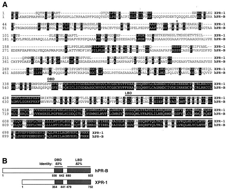
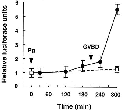
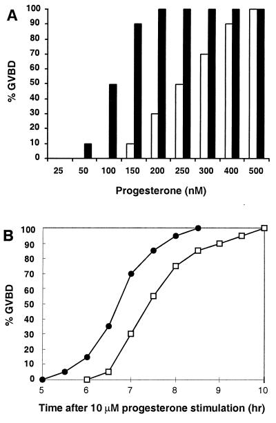
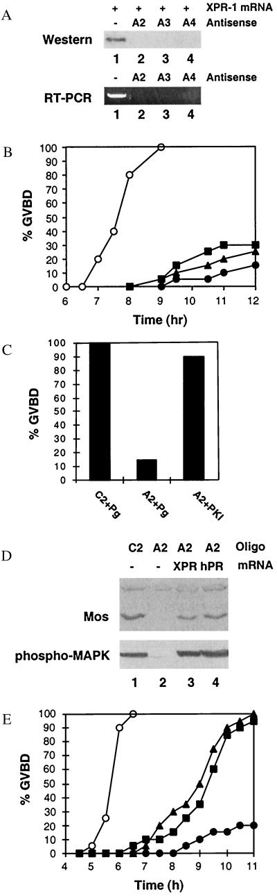
Quiescent full-grown Xenopus oocytes remain arrested at the G(2)/M border of meiosis I until exposed to progesterone, their natural mitogen. Progesterone triggers rapid, nontranscriptional responses that lead to the translational activation of stored mRNAs, resumption of the meiotic cell cycles, and maturation of the oocyte into a fertilizable egg. It has long been presumed that progesterone activates the oocyte through a novel nontranscriptional signaling receptor. Here, we provide evidence that a conventional transcriptional progesterone receptor cloned from Xenopus oocytes, XPR-1, is required for oocyte activation. Overexpression of XPR-1 through mRNA injection increases sensitivity to progesterone and accelerates progesterone-activated cell cycle reentry. Injection of XPR-1 antisense oligonucleotides blocks the ability of oocytes to respond to progesterone; these oocytes are rescued by subsequent injection of XPR-1 or the human progesterone receptor PR-B. Antisense-treated oocytes can be activated in response to inhibition of protein kinase A, one of the earliest known changes occurring downstream of progesterone stimulation. These results argue that the conventional progesterone receptor also functions as the signaling receptor that is responsible for the rapid nontranscriptional activation of frog oocytes.


Figures

Figure 1
Figure 1
Amino acid sequence and domain structure of XPR-1. (A) Predicated protein sequence of XPR-1 and alignment with hPR-B (GenBank accession no. M15716). Identical residues are shaded. Regions comprising the DBD and LBD are indicated by lines above the sequences. * indicates the start of the A form of hPR; −, alignment gap. (B) Comparison of XPR-1 and hPR-B domain structure. Percent sequence identities in the LBD and DBD are indicated.
Figure 2
Figure 2
XPR-1-regulated transcriptional activation of a luciferase reporter construct. Oocytes were either injected with 1 ng of XPR-1 mRNA (●) or left uninjected (□) and incubated for 16 h. Then, 3 ng of luciferase reporter construct DNA (PRE2-tata-Luc) was injected into the nucleus of each oocyte and cells were stimulated with 10 μM progesterone. At the indicated times, groups of 10 injected oocytes were collected and assayed for luciferase activity as described in Materials and Methods. Similar results were seen in two separate experiments.
Figure 3
Figure 3
Effects of overexpressing XPR-1 in oocytes. (A) Sensitivity to progesterone. Oocytes were injected with H2O (□) or 1 ng of XPR-1 mRNA (■), incubated overnight, and then stimulated with progesterone at indicated concentrations. Cells were scored for GVBD after 16 h. Ten oocytes from the same unprimed frog were tested at each concentration of hormone, because the EC50 varied significantly from frog to frog. The relative increase in sensitivity to progesterone was similar in three different experiments. (B) Kinetics of GVBD. Groups of 40 oocytes from unprimed females were injected with H2O (□) or 1 ng of XPR-1 mRNA (●), incubated overnight, and then stimulated with 10 μM progesterone. GVBD was scored at indicated times. Similar results were seen in four separate experiments.
Figure 4
Figure 4
Effect of antisense ablation of XPR-1. (A) Injection of XPR-1 antisense oligonucleotides blocks synthesis of injected XPR-1 mRNA and ablates endogenous XPR-1 mRNA. (Upper) Oocytes were injected with 1 ng of XPR-1 mRNA in 1× injection buffer (25 nl) or XPR-1 mRNA plus individual antisense oligos A2, A3, or A4 at 25 ng per oocyte at 1 ng/nl. After 16 h of incubation, the expression levels of XPR-1 protein from two oocytes per condition were determined on immunoblots using antibody against hPR-B (C-19; Santa Cruz Biotechnology). Lane 1, buffer-injected; lane 2, injected with A2; lane 3, injected with A3; lane 4, injected with A4. (Lower) Endogenous XPR-1 mRNA levels were determined by nested RT-PCR, as described in Materials and Methods. Lanes are as in Upper. (B) Effect of antisense oligonucleotides on GVBD. Oocytes were injected with 25 nl of buffer (○) or 1 ng/nl individual antisense oligonucleotides (●, A2; ▴, A3; ■, A4). After 8 days of incubation, 20 healthy-looking oocytes from each group were stimulated with 1.5 μM progesterone and scored for GVBD at the indicated times. Similar results were obtained in five separate experiments. The effects of antisense oligos were obvious by day 4. Control oligos did not delay GVBD (not shown). (C) Effects of the PKA inhibitor PKI on XPR-1-depleted oocytes. Six days after injection of antisense oligonucleotide A2 or its control oligonucleotide C2, oocytes were either stimulated with 1.5 μM progesterone or injected with 10 nl of PKI (9 units/μl) in the absence of progesterone and GVBD was scored after 16-h incubation. (D) mos synthesis and MAP kinase activation. Oocytes were injected with 25 ng of control (C2) or antisense (A2) oligonucleotides and incubated for 4 days. Groups of A2-injected oocytes were then injected again with 20 ng of XPR-1 or hPR-B mRNA as indicated and incubated for another 2 days. Finally, oocytes were treated with 1.5 μM progesterone for 16 h. At 16 h, three oocytes from each group were lysed and immunoblotted with antibodies against mos or phospho-MAP kinase. (E) Rescue of XPR-1-depleted oocytes by injection of XPR-1 or hPR-B mRNA. Oocytes were injected with 25 ng of control (C2) or antisense (A2) oligonucleotides, incubated for 4 days, and then injected with either XPR-1 or hPR-B mRNA as indicated. After 36 h, oocytes were stimulated with 1.5 μM progesterone and GVBD was scored at the indicated times. ○, 25 ng of C2; ●, 25 ng of A2; ■, 25 ng of A2 plus 25 ng of XPR-1 mRNA; ▴, 25 ng of A2 plus 25 ng of hPR-B mRNA.

Comment in

References

    1. Masui Y, Shibuya E K. In: Molecular Regulation of Nuclear Events in Mitosis and Meiosis. Schlegel R A, Halleck M S, Rao P N, editors. New York: Academic; 1987. pp. 1–42.
    1. Cork R J, Robinson K R. Zygote. 1994;2:289–299. - PubMed
    1. Maller J L. Biol Cell. 1998;90:453–460. - PubMed
    1. Ferrell J E. BioEssays. 1999;21:833–842. - PubMed
    1. Xu L, Glass C K, Rosenfeld M G. Curr Opin Genet Dev. 1999;9:140–147. - PubMed

Publication types

MeSH terms

Associated data