. 2000 Nov;13(5):599-609.
doi: 10.1016/s1074-7613(00)00060-1.

Control of B cell production by the adaptor protein lnk. Definition Of a conserved family of signal-modulating proteins

Affiliations

Control of B cell production by the adaptor protein lnk. Definition Of a conserved family of signal-modulating proteins

S Takaki et al. Immunity. 2000 Nov.

Abstract

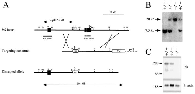
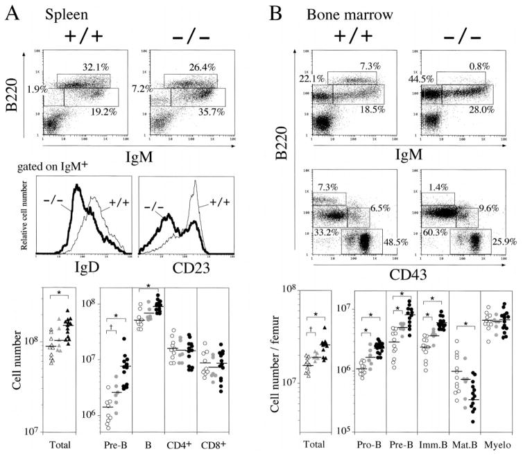
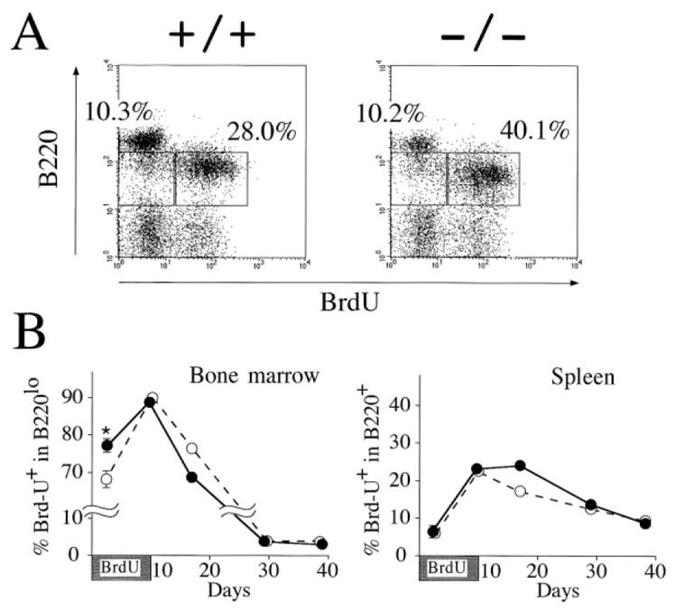
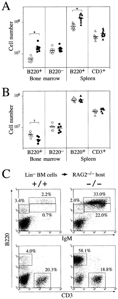
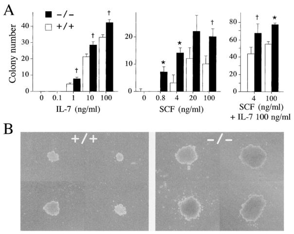
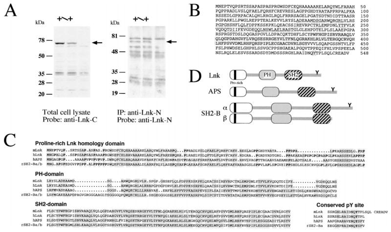
Lnk is an SH2 domain-containing adaptor protein expressed preferentially in lymphocytes. To illuminate the importance of Lnk, we generated lnk(-/-) mice. Whereas T cell development was unaffected, pre-B and immature B cells accumulated in the spleens. In the bone marrow, B-lineage cells were proportionately increased, reflecting enhanced production of pro-B cells that resulted in part from hypersensitivity of precursors to SCF, the ligand for c-kit. Hence, Lnk ordinarily acts to regulate B cell production. Further characterization of lnk(-/-) mice also revealed that full-length Lnk is a 68 kDa protein containing a conserved proline-rich region and a PH domain. Lnk is a representative of a multigene adaptor protein family whose members act, by analogy with Lnk, to modulate intracellular signaling.


Figures

Figure 1
Figure 1. Targeted Disruption of the Murine lnk Gene
(A) Targeting vector and homologous recombination at the lnk locus. Top: a partial restriction map of the murine lnk locus. Known exons are depicted as black boxes. Restriction sites for BamHI (B), BglII (Bg), and EcoRI (E) are indicated, as are the external and cDNA probe. Middle: the lnk gene targeting construct. A BglII fragment containing most of the reported coding sequence of lnk with a neomycin phosphotransferase cassette (Neo). Bottom: predicted structure of the disrupted lnk gene. The exon containing the true initiation codon identified in this study (see Results) is indicated by asterisks. (B) Representative Southern blot analysis of genomic DNA of progeny mice born from a heterozygote cross. DNA was digested with BglII and probed with the external probe. (C) Expression of the lnk transcript in spleen. Poly(A)+ RNA (3 μg) from splenocytes was separated, transferred, and hybridized with mouse lnk cDNA probe (upper) or β-actin probe (lower).
Figure 2
Figure 2. Accumulation of B-Lineage Cells in lnk−/− Mice
(A) Splenic B cells are increased and show immature phenotypes in lnk−/− mice. Representative two-color fluorescence plots showing expression of B220 and IgM (upper panels) on splenocytes and expression of IgD (middle left) or CD23 (middle right) on IgM+ B cells. Percentages represent the fractions of the total gated live cells that fall into the indicated boxes. Cellularity of pre-B (B220+IgM), B (B220+IgM+), CD4+ T, and CD8+ T in spleen (lower panels). lnk+/+, lnk+/−, and lnk−/− mice are represented by open, shaded, and closed symbols, respectively. *p < 0.01, †p < 0.05 by the Student’s t test. (B) B cell precursors are accumulated in the bone marrow of lnk−/− mice. Representative two-color fluorescence plots showing expression of B220 and IgM (upper panels), as well as expression of B220 and CD43 (middle panels) on bone marrow cells. Percentages represent the fractions of the total gated live cells within the indicated boxes. Cellularity of pro-B: B220+CD43+, pre-B: B220+CD43IgM, immature B: B220loIgM+, mature B: B220hiIgM+, myeloid: B220CD43+ in bone marrow (lower panels). lnk+/+, lnk+/−, and lnk−/− mice are represented by open, shaded, or closed symbols, respectively. *p < 0.01, †p < 0.05.
Figure 3
Figure 3. In Vivo Incorporation of BrdU by lnk−/− B Cell Precursors and Splenic B Cells
(A) Mice were fed BrdU in drinking water for 40 hr, and bone marrow cells were stained for B220 expression and BrdU content. Percentages represent the fractions of the gated lymphocytes that fall into the indicated boxes. The data shown are representative results from three independent experiments. (B) Mice were fed BrdU for 10 days. Two lnk−/− (closed circles) and lnk−/− (open circles) were killed and analyzed at each time point (except day 2). The mean percentage of BrdU+ cells in B220lo bone marrow cells (left) or the mean percentage of BrdU+ cells in B220+ splenocytes (right) is shown. At day2, 4 lnk−/− and 4 lnk+/+ mice were analyzed and the mean ± SEM is shown. *p < 0.01 compared with +/+ mice.
Figure 4
Figure 4. Defects in B Cell Precursors Are Responsible for B Cell Overproduction
(A) Irradiated C57BL/6 mice were transferred with bone marrow cells from lnk+/+ (open symbols) or lnk−/− (closed symbols). The resulting chimeric mice were analyzed at 7–8 weeks after bone marrow transfer. Data shown are cell numbers of indicated cell populations in the bone marrow (per femur) or the spleen. *p < 0.01. (B) lnk+/+ (open symbols) or lnk−/− mice (closed symbols) were irradiated and transferred with C57BL/6 bone marrow cells. †p < 0.05. (C) Successful repopulation of lnk−/− precursor cells into B cell compartment in a competitive environment. Bone marrow cells negative for the lineage makers (Lin; B220, CD3ε, Gr-1, Mac-1, and TER-119) were isolated from lnk−/− or lnk+/+ mice and were injected (2 × 10⁵ per head) into RAG2−/− mice without irradiation. Splenocytes from the injected mice were analyzed at 7–8 weeks after injection. Displayed are representative two-color fluorescence plots showing expression of IgM and B220 (upper panels) or expression of B220 and CD3 (lower panels). Percentages represent the fractions of the total gated live cells that fall into the indicated boxes. The data shown are representative results from three independent experiments.
Figure 5
Figure 5. Colony Formation by Bone Marrow Cells in Response to IL-7 and SCF
(A) The numbers of colonies appearing in clonal cultures of bone marrow cells in semi-solid medium containing various concentrations of IL-7 (left) or SCF (middle) or containing both SCF and IL-7 (right) were compared between lnk+/+ (open bars) and lnk−/− (solid bars) mice. The values are the mean colony number (± SD) of triplicate cultures. *p < 0.005, †p < 0.05 compared with +/+ mice. The data shown are representative results from two independent experiments. (B) Lymphoid colonies formed in the presence of SCF (4 ng/ml) and IL-7 (100 ng/ml).
Figure 6
Figure 6. Revised Structure of Lnk and Its Family Members
(A) Western blot analysis of splenocytes by anti-Lnk antibodies. Splenocyte lysates prepared from lnk+/+, lnk+/−, or lnk−/− mice were analyzed by immunoblotting probed with anti-Lnk-C-terminal antibodies (left panel). Immunoprecipitates from splenocyte lysates by anti-Lnk-N-terminal antibodies were immunoblotted using biotinylated anti-Lnk-N-terminal antibodies directed against a newly identified N-terminal portion of full-length Lnk protein (right panel). Arrows indicate a 68 kDa band representing Lnk. (B) Amino acid sequence of mouse Lnk predicted from a combined sequence of lnk cDNAs. N-terminal region rich with proline residues and homologous to APS and SH2-B (dotted line), PH domain (solid line), SH2 domain (shaded), a conserved tyrosine phosphorylation site at the C-terminal end (boxed) are indicated. (C) Amino acid sequence alignments of proline-rich N-terminal region, PH domain, SH2 domain, and conserved tyrosine phosphorylation site. Amino acid residues identical in at least two of three mammalian Lnk family members (Lnk, APS, and SH2-B) are indicated by shaded boxes. Proline residues in the N-terminal region are highlighted by boldface. (D) Schematic representation of Lnk-family members.

References

    1. Ahmed Z, Smith BJ, Kotani K, Wilden P, Pillay TS. APS, an adapter protein with a PH and SH2 domain, is a substrate for the insulin receptor kinase. Biochem J. 1999;341:665–668. - PMC - PubMed
    1. Akasaka T, Tsuji K, Kawahira H, Kanno M, Harigaya K, Hu L, Ebihara Y, Nakahata T, Tetsu O, Taniguchi M, Koseki H. The role of mel-18, a mammalian Polycomb group gene, during IL-7-dependent proliferation of lymphocyte precursors. Immunity. 1997;7:135–146. - PubMed
    1. Alberola-Ila J, Takaki S, Kerner JD, Perlmutter RM. Differential signaling by lymphocyte antigen receptors. Annu Rev Immunol. 1997;15:125–154. - PubMed
    1. Appleby MW, Gross JA, Cooke MP, Levin SD, Qian X, Perlmutter RM. Defective T cell receptor signaling in mice lacking the thymic isoform of p59fyn. Cell. 1992;70:751–763. - PubMed
    1. Baird AM, Gerstein RM, Berg LJ. The role of cytokine receptor signaling in lymphocyte development. Curr Opin Immunol. 1999;11:157–166. - PubMed

Publication types

Substances

Associated data

LinkOut - more resources