. 2000 Dec 1;14(23):2976-88.
doi: 10.1101/gad.188700.

FtsK functions in the processing of a Holliday junction intermediate during bacterial chromosome segregation

Affiliations

FtsK functions in the processing of a Holliday junction intermediate during bacterial chromosome segregation

F X Barre et al. Genes Dev. .

Abstract

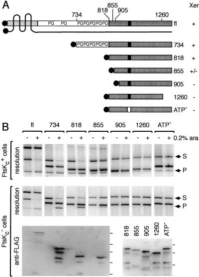
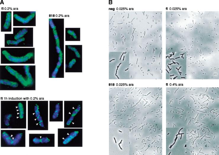
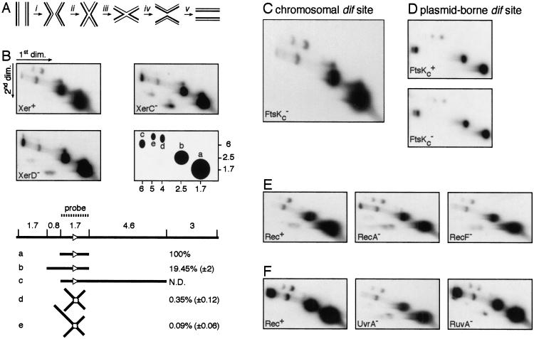
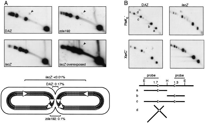
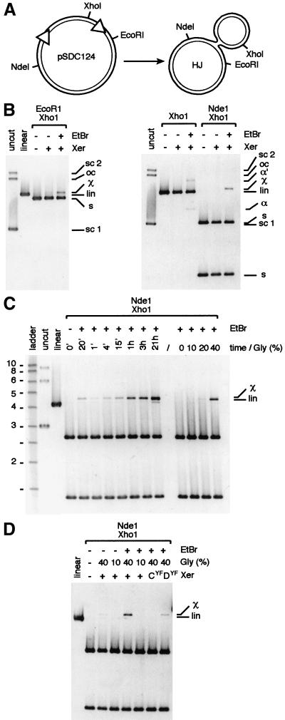
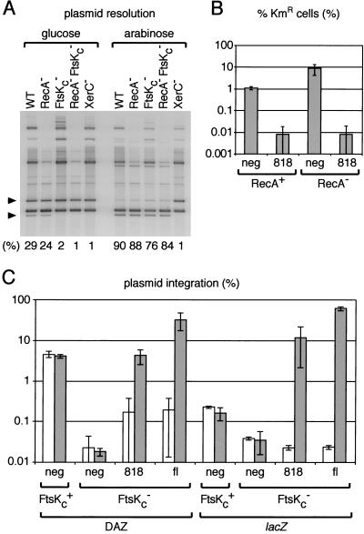
In bacteria with circular chromosomes, homologous recombination can generate chromosome dimers that cannot be segregated to daughter cells at cell division. Xer site-specific recombination at dif, a 28-bp site located in the replication terminus region of the chromosome, converts dimers to monomers through the sequential action of the XerC and XerD recombinases. Chromosome dimer resolution requires that dif is positioned correctly in the chromosome, and the activity of FtsK, a septum-located protein that coordinates cell division with chromosome segregation. Here, we show that cycles of XerC-mediated strand exchanges form and resolve Holliday junction intermediates back to substrate irrespective of whether conditions support a complete recombination reaction. The C-terminal domain of FtsK is sufficient to activate the exchange of the second pair of strands by XerD, allowing both intra- and intermolecular recombination reactions to go to completion. Proper positioning of dif in the chromosome and of FtsK at the septum is required to sense the multimeric state of newly replicated chromosomes and restrict complete Xer reactions to dimeric chromosomes.


Figures

Figure 1
Figure 1
Minimal region of FtsK required for Xer recombination. (A) FtsK derivatives used in this study. The Nterminal domain of FtsK, domain 1, is represented by a lightly shaded box, and the four putative transmembrane regions are shown as black lines crossing the protein. The extreme N terminus points toward the cytoplasm, as well as domain 2 and 3 of the protein. The C-terminal domain, domain 3, is shown as a dark box, and the putative nucleotide binding site as a black rectangle. The Flag peptide is shown as shaded circles. The N-terminal (734, 818, 855, 905) and C-terminal (1260) amino acid of each protein are indicated on top of the full-length protein (fl). The relative ability of each protein to support Xer recombination on plasmids is indicated by +/− signs on the right of the diagram. (B) Xer recombination between plasmid-borne dif sites in cells expressing FtsK deletions. FtsKc+ (DS941) and FtsKc (DS9041) cells (as shown to the left of the gels) were transformed with the different ftsK expression vectors, as indicated along the top of the gels. Plasmid-containing cells were grown for 5 h in arabinose (+, 0.2% ara) or in glucose (−). Recombination between the two dif sites carried by the substrate (S) leads to the formation of two smaller product circles, of which one can replicate (P). The substrate and product bands are indicated by arrows to the right of the gels. The third band present in each lane is the ftsK expression vector. Expression of the FtsK derivatives was verified by Western blot analysis after resolution of the proteins on a 6% (fl, 734, 818, 855) or a 8% (818, 855, 905, 1260, ATP) SDS-PAGE. Complete repression (−) was verified for fl, 734, 818, and 855. The position of the protein molecular weight markers are indicated on the right of each blot and correspond from bottom to top to 32, 47, 67, 83, and 175 kD. The relatively low signal with fl may reflect a poorer transfer to the membrane.
Figure 2
Figure 2
(A) Cellular distribution of fl and 818 FtsK proteins. Induction with arabinose (0.2%) was for ∼25 generation in FtsKc (DS9041) cells (top) or for 1 h in FtsKc+ (DS941) cells (bottom). The localization of the DNA and of the FtsK proteins is shown in blue and green pseudocolors, respectively. White arrows indicate septum ring-like structures. (B) Morphology of FtsKc cells expressing various amounts of fl or 818 FtsK proteins or carrying a control expression vector (neg).
Figure 3
Figure 3
In vivo detection of HJ intermediates at the chromosomal dif site. (A) The different steps of Xer site-specific recombination. (i) Synapsis; (ii) first pair of strand exchanges; (iii) HJ conformational change; (iv) second pair of strand exchanges; (v) dissociation. (B) Detection of HJs at dif in Xer+ (AB1157), XerC (DS8029), and XerD (GR18) cells. EcoRV was used to restrict the DNA samples. A simplified scheme of the hybridization signals is shown along with molecular weights in kilobase pairs. Five spots were detected that correspond to expected linear (a, b, and c) or joint (d and e) molecules. The EcoRV restriction digest map of the dif region and the molecules that can be attributed to the a, b, c, d, and e spots are represented below the blots. The dif site is shown as a white triangle. The relative intensity of each spot to the intensity of spot (a) was calculated in XerC+ XerD+ cells. Numbers correspond to the mean and standard deviation of values from eight independent experiments. (C) HJs at the chromosomal dif site in FtsKc cells (MA3). (D) HJs in FtsKc+ (AB1157) and FtsKc cells carrying a multicopy plasmid with a single dif site. (E) HJs at the chromosomal dif site in Rec+ (AB1157), RecA (GR19), and RecF (DS941) cells. (F) HJs at the chromosomal dif site in Rec+ (JJC40), UvrA (N3137), and RuvA (JJC662) cells. The smeary bands below the arc of linear DNA in some of the gels are blotting artefacts and contain small amounts of the major DNA species from the linear arc.
Figure 4
Figure 4
Effect of the location of dif on the level of HJs. (A) HJs at dif (indicated by arrowheads) were compared in three strains carrying a single site in DAZ (LN3080), at 150 kb to the left side of DAZ (zda192, LN3577) or at 1 Mb to the right side of DAZ (lacZ, LN3091). The positions of the two sister dif sites in dividing cells are represented below the blots. Numbers indicate the relative intensity of the HJ spot to the intensity of the smaller linear fragment (mean of two independent experiments). (B) Increased and location-independent levels of HJ in cells carrying two tandem dif sites. HJ levels were compared in FtsKc and XerC cells carrying a dif-Km-dif cassette in DAZ or in lacZ (FC313, FC354, FC235, FC284). EcoRV (E) and HindIII (H) were used to restrict DNA. The restriction digest map of the dif-Km-dif cassette and the molecules that can be attributed to the a, b, c, and d spots are represented below the blots. Note that the intensity of spot d must be compared with the sum of the intensities of spot a and b.
Figure 5
Figure 5
In vitro HJ formation by XerCD on a supercoiled plasmid carrying two dif sites. (A) restriction map of plasmid pSDC124 and of the HJ created by action of XerCD. (B) One-hour reactions in 40% glycerol. (C) Time course experiment in 40% glycerol and effect of the glycerol concentration in 1-h reactions. Molecular weights are in kilobase pairs. (D) Determination of the Xer recombinase whose catalytic activity is necessary for HJ formation. One-hour reactions in 40% glycerol used catalytically inactive mutants of XerC (CYF) and XerD (DYF) mixed with their functional partner. pSCD124 was left untreated (uncut, linear) or treated with the Xer recombinases (Xer). EtBr was added at the end of reactions as indicated. For the 20′ time point, EtBr was added at the beginning of the reaction. sc 1, sc 2, lin, and oc, supercoiled monomer, supercoiled dimer, linear, open circle forms of pSDC124, respectively. s, restriction fragments from pSCD124; χ, χ-molecule containing a HJ; α and α′, supercoiled and nicked α-molecules containing a HJ.
Figure 6
Figure 6
Overexpression of FtsKc abolishes the requirement for homologous recombination and for the location of dif in DAZ. (A) Plasmid resolution assay in WT (AB1157), RecA (GR19), FtsKc (MA3), RecA FtsKc (GR59), and XerC (DS8029) cells under conditions of repression (glucose) or induction (0.4% arabinose) of 818 expression. The bands corresponding to the supercoiled substrate and supercoiled replicative product are indicated by the top and bottom black triangle, respectively. Numbers indicate the proportion of supercoiled product to total supercoiled substrate in each lane. (B) dif-Km-dif cassette resolution assay. RecA+ (FC235) and RecA (FC217) cells were transformed with a control expression vector (neg) or with the vector expressing the 818 FtsK derivative. They then were transformed with pFC225, which carries a functional xerC gene. The proportion of kanamycin resistant cells in the cultures (KmR) was quantitated after overnight growth in the presence of 0.2% arabinose. Results from a typical experiment are shown. (C) Plasmid integration assay. FtsKc+ and FtsKc cells carrying a single chromosomal dif site in DAZ or in lacZ (DS941, NL250, DS9041, MA2) were transformed with a control expression vector (neg), with the 818 expression vector or with the fl expression vector. They then were transformed with pFH127, which carries a single dif site. The proportion of cells having integrated this plasmid was quantitated after overnight growth at 30°C in the presence of glucose (white bars) or 0.2% arabinose (gray bars). Shown is the result of at least two independent experiments.

References

    1. Arciszewska LK, Sherratt DJ. Xer site-specific recombination in vitro. EMBO J. 1995;14:2112–2120. - PMC - PubMed
    1. Arciszewska LK, Baker RA, Hallet B, Sherratt DJ. Coordinated control of XerC and XerD catalytic activities during Holliday junction resolution. J Mol Biol. 2000;299:391–403. - PubMed
    1. Austin S, Ziese M, Sternberg N. A novel role for site-specific recombination in maintenance of bacterial replicons. Cell. 1981;25:729–736. - PubMed
    1. Barre FX, Ait-Si-Ali S, Giovannangeli C, Luis R, Robin P, Pritchard LL, Helene C, Harel-Bellan A. Unambiguous demonstration of triple-helix-directed gene modification. Proc Natl Acad Sci. 2000;97:3084–3088. - PMC - PubMed
    1. Begg KJ, Dewar SJ, Donachie WD. A new Escherichia coli cell division gene, ftsK. J Bacteriol. 1995;177:6211–6222. - PMC - PubMed

Publication types

MeSH terms

LinkOut - more resources