. 2000 Dec 1;14(23):3065-73.
doi: 10.1101/gad.182100.

Role of Bak in UV-induced apoptosis in skin cancer and abrogation by HPV E6 proteins

Affiliations

Role of Bak in UV-induced apoptosis in skin cancer and abrogation by HPV E6 proteins

S Jackson et al. Genes Dev. .

Abstract

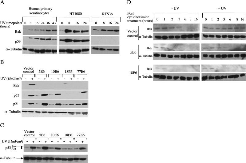
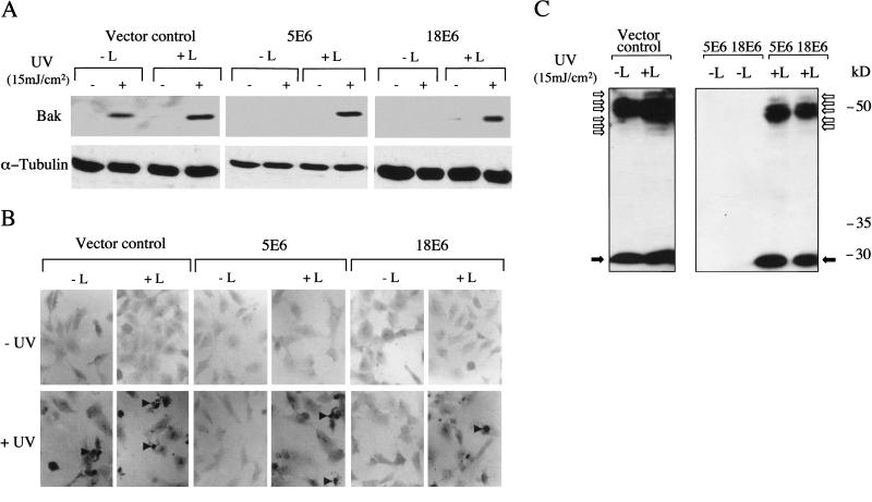
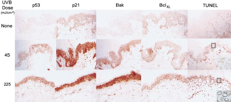
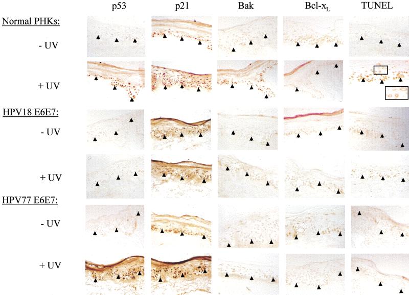
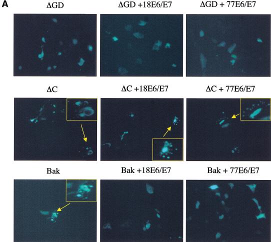
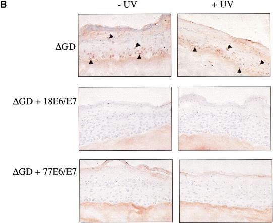
Ultraviolet B (UVB) damage is recognized as the most important etiological factor in the development of skin cancer. Human papillomaviruses (HPV) have also been implicated in the disease, although the mechanism of action of these viruses remains unknown. We present evidence here that Bak protein is involved in signaling apoptosis in the skin in response to UVB damage, and that cutaneous HPV E6 proteins target and abrogate Bak function by promoting its proteolytic degradation both in vitro and in regenerated epithelium. Additionally, HPV positive skin cancers had undetectable levels of Bak in contrast to HPV negative cancers, which expressed Bak. This study supports a link between the virus and UVB in the induction of HPV-associated skin cancer and reveals a survival mechanism of virally infected cells.


Figures

Figure 1
Figure 1
Induction of Bak by UVB and abrogation of the Bak pathway by E6 proteins. (A) Human primary keratinocytes, HT1080 cells and human p53-null keratinocytes (RTS3b line) were treated with UVB (15 mJ/cm2) and the cells harvested at 0–43 h (only human primary keratinocytes survived after 24 h). Equal protein samples were resolved by SDS-PAGE, with Bak, p53 and α-tubulin levels detected by Western blotting using monoclonal antibodies. (B) HT1080 E6 polyclonal cell lines were treated with UVB (15 mJ/cm2) and the cells harvested 24 h later. Proteins were detected as in A. The membrane was exposed to X-ray film for 1 min prior to development. (C) Polymorphic forms of p53 from B were resolved on a 10% PAG and detected as previously. (D) HT1080 E6 cells were treated with 60 μg/mL cycloheximide 5 min post UV treatment (15 mJ/cm2). Protein extracts were prepared at the indicated time points following cycloheximide treatment and Bak levels determined by Western blotting. The membrane was exposed to X-ray film for 20 min prior to development to detect low levels of Bak.
Figure 2
Figure 2
Ubiquitin-dependent proteolytic degradation of Bak and induction of apoptosis following UV treatment. (A) Western blot analysis of endogenous Bak levels in HT1080 vector control cells, 5E6 cells and 18E6 cells grown in the presence (+L) and absence (−L) of the proteasome inhibitor lactacystin (5 μM) with (+) and without (−) UVB (15 mJ/cm2) treatment. Lactacystin was added 2 h prior to the 24-h cell harvesting timepoint. (B) Parallel cultures of cells seeded onto glass coverslips were treated with UV and lactacystin as in A. Cells were fixed in paraformaldehyde and apoptotic cells detected by TUNEL (Magnification, 200×). (C) HT1080 cells were exposed to UVB (15 mJ/cm2) and were grown in the presence (+L) and absence (−L) of lactacystin for 2 h prior to cell harvesting, as in A. Cells were lysed 24 h post UVB in SDS sample buffer diluted in RIPA buffer containing iodoacetamide (10 mM), ubiquitinated Bak was detected by Western blotting.
Figure 3
Figure 3
Bak induction in normal skin by UVB. Skin specimens were irradiated with 45 or 225 mJ/cm2 UVB and snap frozen 24 h posttreatment for immunohistochemical analysis. Apoptotic cells were detected using TUNEL (original magnification, 100×, insert, 400×).
Figure 4
Figure 4
Effects of irradiation on HPV18 and 77 E6/E7 transfected keratinocytes grown in organotypic culture. Endogenous p53, p21, Bak and Bcl-xL protein levels were detected by immunohistochemistry in HPV 18 and 77 E6/E7 cultures 24 h post UV treatment (45 mJcm-2) and compared to control PHK raft cultures (original magnification, 100×).
Figure 5
Figure 5
Inhibition of Bak apoptotic activity but not degradation is required to regenerate epidermis in vitro. (A) Normal keratinocytes were transfected with either Bak or the ΔGD or ΔC mutants either alone or together with HPVE6/E7. EGFP was used as a marker of transfection. 16 h post-transfection the cells were monitored for morphological signs of apoptosis (membrane blebbing, nuclear condensation and fragmentation) (Magnification, 200×; inserts, 400×). (B) ΔGD and ΔGD/E6/E7 cells were seeded onto de-epidermalized dermis and allowed to reform an epithelium. The cells were irradiated with 45 mJcm−2 UVB, harvested 8 h posttreatment and processed for immunohistochemistry. Sections were counterstained with haematoxylin. Positively stained cells are indicated by arrowheads. (Magnification, 200×)
Figure 5
Figure 5
Inhibition of Bak apoptotic activity but not degradation is required to regenerate epidermis in vitro. (A) Normal keratinocytes were transfected with either Bak or the ΔGD or ΔC mutants either alone or together with HPVE6/E7. EGFP was used as a marker of transfection. 16 h post-transfection the cells were monitored for morphological signs of apoptosis (membrane blebbing, nuclear condensation and fragmentation) (Magnification, 200×; inserts, 400×). (B) ΔGD and ΔGD/E6/E7 cells were seeded onto de-epidermalized dermis and allowed to reform an epithelium. The cells were irradiated with 45 mJcm−2 UVB, harvested 8 h posttreatment and processed for immunohistochemistry. Sections were counterstained with haematoxylin. Positively stained cells are indicated by arrowheads. (Magnification, 200×)

References

    1. Allday M, Inman G, Crawford D, Farrell P. DNA damage in human B cells can induce apoptosis, proceeding from G1/S when p53 is transactivation competent and G2/M when it is transactivation defective. EMBO J. 1995;14:4994–5005. - PMC - PubMed
    1. Arends M, McGregor A, Wyllie A. Apoptosis is inversely related to necrosis and determines net growth in tumours bearing constitutively expressed myc, ras, and HPV oncogenes. Am J Path. 1994;144:1045–1057. - PMC - PubMed
    1. Burger H, Hooter K, Boersma AWM, Kortland CJ, Van Den Berg AP, Stoter G. Expression of p53, p21Waf/Cip, Bcl-2, Bax, Bcl-X and Bak in radiation-induced apoptosis in testicular germ cell tumour lines. Int J Radiation Oncology Biol Phys. 1998;41:415–424. - PubMed
    1. Chittenden T, Flemington C, Houghton AB, Ebb RG, Gallo GJ, Elangovan D, Chinnadurai G, Lutz RJ. A conserved domain in Bak, distinct from BH1 and BH2, mediates cell death and protein binding functions. EMBO J. 1995;14:5589–5596. - PMC - PubMed
    1. Craiu A, Gaczynska M, Akopian T, Gramm CF, Fenteany G, Goldberg AL, Rock KL. Lactacystin and clasto-lactacystin β-lactone modify multiple proteosome β-subunits and inhibit intracellular protein degradation and major histocompatibility complex class I antigen presentation. J Biol Chem. 1997;272:13437–13445. - PubMed

Publication types

MeSH terms

Substances