. 2000 Dec;124(4):1728-38.
doi: 10.1104/pp.124.4.1728.

Aux/IAA proteins are phosphorylated by phytochrome in vitro

Affiliations

Aux/IAA proteins are phosphorylated by phytochrome in vitro

A Colón-Carmona et al. Plant Physiol. 2000 Dec.

Abstract

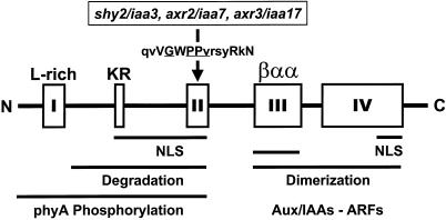
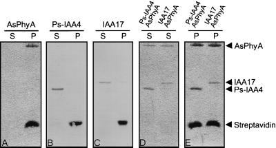
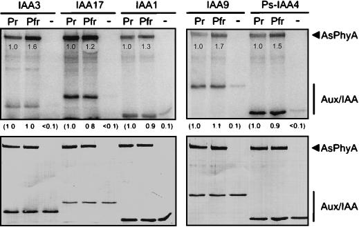
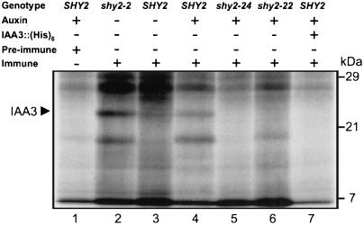
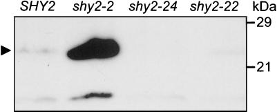
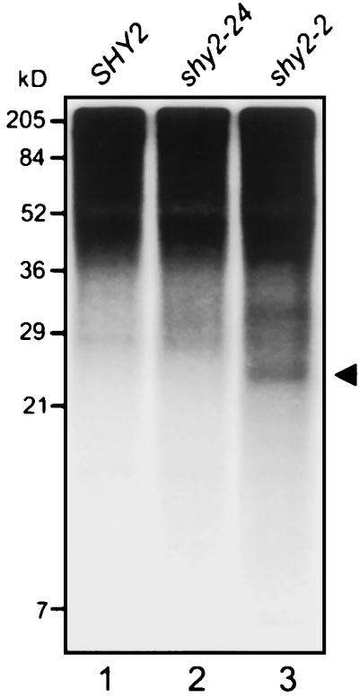
Auxin/indole-3-acetic acid (Aux/IAA) genes encode short-lived transcription factors that are induced as a primary response to the plant growth hormone IAA or auxin. Gain-of-function mutations in Arabidopsis genes, SHY2/IAA3, AXR3/IAA17, and AXR2/IAA7 cause pleiotropic phenotypes consistent with enhanced auxin responses, possibly by increasing Aux/IAA protein stability. Semidominant mutations shy2-1D, shy2-2, axr3-1, and axr2-1 induce ectopic light responses in dark-grown seedlings. Because genetic studies suggest that the shy2-1D and shy2-2 mutations bypass phytochrome requirement for certain aspects of photomorphogenesis, we tested whether SHY2/IAA3 and related Aux/IAA proteins interact directly with phytochrome and whether they are substrates for its protein kinase activity. Here we show that recombinant Aux/IAA proteins from Arabidopsis and pea (Pisum sativum) interact in vitro with recombinant phytochrome A from oat (Avena sativa). We further show that recombinant SHY2/IAA3, AXR3/IAA17, IAA1, IAA9, and Ps-IAA4 are phosphorylated by recombinant oat phytochrome A in vitro. Deletion analysis of Ps-IAA4 indicates that phytochrome A phosphorylation occurs on the N-terminal half of the protein. Metabolic labeling and immunoprecipitation studies with affinity-purified antibodies to IAA3 demonstrate increased in vivo steady-state levels of mutant IAA3 in shy2-2 plants and phosphorylation of the SHY2-2 protein in vivo. Phytochrome-dependent phosphorylation of Aux/IAA proteins is proposed to provide one molecular mechanism for integrating auxin and light signaling in plant development.


Figures

Figure 1
Figure 1
Structural and functional domains of Aux/IAA proteins. The primary structure conserved in the core of domain II is given above the designated box. Uppercase letters denote invariant amino acid residues. Positions affected by missense gain-of-function mutations of the indicated Aux/IAA genes are underlined. Bars below the structural domains delineate regions important to various protein activities or modifications. These include functionally identified signals for nuclear localization (NLS; Abel and Theologis, 1995) and protein degradation (Worley et al., 2000), as well as regions shown to mediate homo- and heterodimerization (Kim et al., 1997; Morgan et al., 1999) and to be modified by phytochrome A phosphorylation (this study).
Figure 2
Figure 2
Aux/IAA proteins and phytochrome A interact in vitro. Recombinant oat phyA (AsPhyA) and IAA proteins were mixed with streptavidin agarose beads, incubated at room temperature, and treated as described in “Materials and Methods.” Shown are SDS-PAGE resolved proteins of the binding reactions after Coomassie Blue staining for AsPhyA (A) and IAA protein (B and C) control incubations, as well as for the test incubations containing AsPhyA and IAA protein (D and E). S indicates the entire supernatant fraction after the first sedimentation step. P denotes the entire pellet fraction after the third washing step with detergent-containing buffer.
Figure 3
Figure 3
Phytochrome A phosphorylates Aux/IAA proteins in vitro. Recombinant oat phyA (AsPhyA) in the Pr form or Pfr form was used in kinase assays with recombinant IAA proteins as described in “Materials and Methods.” To detect any autophosphorylation of IAA proteins, assays were also performed without AsPhyA. Shown are autoradiographs of the kinase reactions after SDS-PAGE and protein transfer to membranes (top), and Coomassie Blue staining of the transferred proteins (bottom). Amounts of phyA autophosphorylation and of Aux/IAA phosphorylation by phyA are expressed relative to the reactions containing the Pr form of phyA and are given below the phyA protein bands and below the top panels, respectively.
Figure 4
Figure 4
Deletion analysis of Ps-IAA4. Kinase assays were conducted in white light with (+) or without (−) recombinant oat phyA (AsPhyA) using full-length (I-IV) Ps-IAA4 (amino acids 1–189) and Ps-IAA4 deletions, domains I-II (amino acids 1–90), and domains III-IV (amino acids 86–189), as protein substrates. Shown are autoradiographs and Coomassie Blue protein staining of the resolved kinase reactions. The arrowhead and bar indicate the positions of oat phyA and of truncated PS-IAA4 polypeptides, respectively.
Figure 5
Figure 5
Immunoprecipitations with anti-IAA3 of [35S]Met-labeled proteins synthesized in vivo. Etiolated seedlings of the indicated genotypes were metabolically labeled, treated with auxin, and extracted as described in “Materials and Methods.” Shown is the autoradiograph (8 d of exposure) for resolved proteins after immunoprecipitations of the extracts with affinity-purified antibodies to IAA3 (lanes 2–7) or with preimmune serum (lane 1). For each immunoprecipitation reaction, 2.1 × 10⁸ cpm of trichloroacetic acid-precipitable material were used. Incorporation of [35S]Met into trichloroacetic acid-precipitable material was between 31% to 37% of the radiolabel measured in total tissue extracts. The arrowhead indicates a 23-kD protein identified as endogenous IAA3.
Figure 6
Figure 6
Detection of IAA3 proteins by immunoblot analysis. Seedlings of the indicated genotypes were grown for 5 d in darkness and 300 μg of total proteins were resolved by SDS-PAGE, transferred to a membrane, and probed with affinity-purified IAA3 antibodies as described in “Materials and Methods.” Detection by enhanced chemiluminescence was for 20 min of exposure time. The arrowhead indicates a 23-kD protein identified as endogenous IAA3.
Figure 7
Figure 7
Immunoprecipitations of [32P]orthophosphate-labeled IAA3 proteins synthesized in vivo. Etiolated seedlings of the indicated genotypes were radioactively labeled, treated with auxin (wild-type and shy2-24), and extracted as described in “Materials and Methods.” Shown is the autoradiograph of resolved proteins after immunoprecipitations of the extracts with affinity-purified antibodies to IAA3 (10 d of exposure). The arrowhead indicates a 23-kD protein, likely representing IAA3. The labeled protein bands seen in the region above 36 kD are due to non-specific binding to protein A agarose beads (data not shown).

References

    1. Abel S, Nguyen D, Theologis A. The PS-IAA4/5-like family of early auxin-inducible mRNAs in Arabidopsis thaliana. J Mol Biol. 1995;251:533–549. - PubMed
    1. Abel S, Oeller PW, Theologis A. Early auxin-induced genes encode short-lived nuclear proteins. Proc Natl Acad Sci USA. 1994;91:326–330. - PMC - PubMed
    1. Abel S, Theologis A. A polymorphic bipartite motif signals nuclear targeting of early auxin-induced proteins related to PS-IAA4 from pea (Pisum sativum) Plant J. 1995;8:87–96. - PubMed
    1. Abel S, Theologis A. Early genes and auxin action. Plant Physiol. 1996;111:9–17. - PMC - PubMed
    1. Ahmad M, Jarillo JA, Smirnova O, Cashmore AR. The CRY1 blue light photoreceptor of Arabidopsis interacts with phytochrome A in vitro. Mol Cell. 1998;1:939–948. - PubMed

Publication types

MeSH terms