. 2000 Dec;124(4):1800-13.
doi: 10.1104/pp.124.4.1800.

The isolation and characterization in yeast of a gene for Arabidopsis S-adenosylmethionine:phospho-ethanolamine N-methyltransferase

Affiliations

The isolation and characterization in yeast of a gene for Arabidopsis S-adenosylmethionine:phospho-ethanolamine N-methyltransferase

C P Bolognese et al. Plant Physiol. 2000 Dec.

Abstract

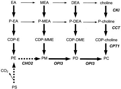
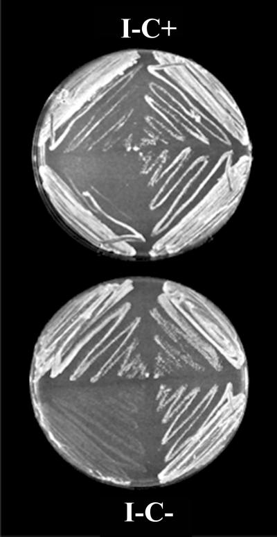
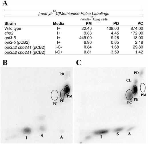
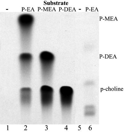
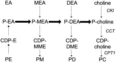
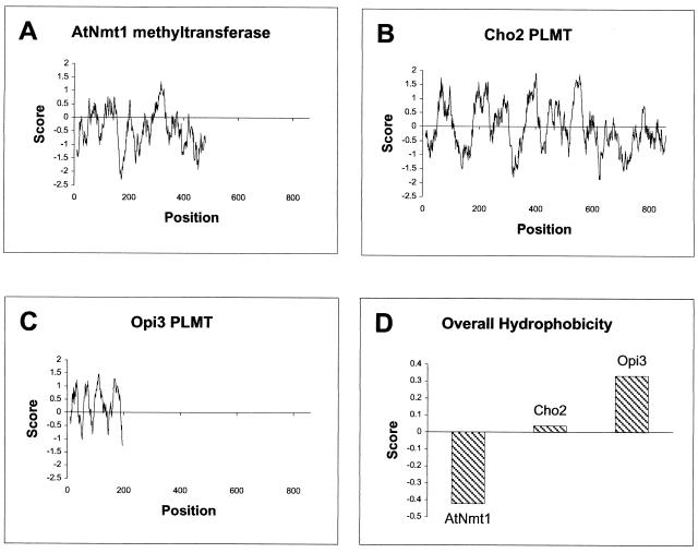
Saccharomyces cerevisiae opi3 mutant strains do not have the phospholipid N-methyltransferase that catalyzes the two terminal methylations in the phosphatidylcholine (PC) biosynthetic pathway. This results in a build up of the intermediate phosphatidylmonomethylethanolamine, causing a temperature-sensitive growth phenotype. An Arabidopsis cDNA library was used to isolate three overlapping plasmids that complemented the temperature-sensitive phenotype. Phospholipid analysis showed that the presence of the cloned cDNA caused a 65-fold reduction in the level of phosphatidylmonomethylethanolamine and a significant, though not equivalent, increase in the production of PC. Sequence analysis established that the cDNA was not homologous to OPI3 or to CHO2, the only other yeast phospholipid N-methyltransferase, but was similar to several other classes of methyltransferases. S-adenosyl-Met:phospho-base N-methyltransferase assays revealed that the cDNA catalyzed the three sequential methylations of phospho-ethanolamine to form phospho-choline. Phospho-choline is converted to PC by the CDP-choline pathway, explaining the phenotype conferred upon the yeast mutant strain by the cDNA. In accordance with this the gene has been named AtNMT1. The identification of this enzyme and the failure to isolate a plant phospholipid N-methyltransferase suggests that there are fundamental differences between the pathways utilized by yeast and by some plants for synthesis of PC.


Figures

Figure 1
Figure 1
PC biosynthesis in yeast and plants. The methyl donor for each methylation step is SAM. Pathways occurring only in yeast are dashed lines; pathways found in yeast and plants are large arrows; plant pathways are small arrows. Adapted from Moore (1993) and McGraw and Henry (1989). PS, Phosphatidylserine.
Figure 2
Figure 2
Sequence analysis. A, Partial restriction map of the complementing cDNAs. pCB1 is approximately 1,800 bp, pCB2 is approximately 2,000 bp, and pCB3 is approximately 4,000 bp. N, NotI; B, BgIII; H, HindIII; S, SacI; Inset, pFL61. B, Genomic organization of AtNMT1. The AtNMT1 gene has 11 introns. Boxes denote exons 1 through 12. The coding sequence is 1,673 bp.
Figure 3
Figure 3
AtNMT1 nucleotide sequence. Shown is the nucleotide sequence of the AtNMT1 gene (complete cDNA sequence GenBank accession no. is AF197940) and the deduced amino acid sequence using the one-letter abbreviations for the amino acids. The boxed sequences of the AtNMT1 gene product are two putative SAM-binding domains. The first domain is in the N-terminal portion of the protein from amino acid 57 to 156. Within the SAM-binding domain, there are four motifs: Motif I (positions 57–65), post-I (positions 79–82), Motif II (positions 118–126), and Motif III (positions 147–156). The second putative SAM-binding domain is in the C-terminal portion of the protein from amino acid 286 to 383: Motif I (positions 286–294), post-I (positions 308–312), Motif II (positions 347–354), and Motif III (positions 374–383). The bold letters in the nucleotide sequence denote the intron/exon boundary.
Figure 4
Figure 4
Complementation analysis of Opi3 phenotypes. A, The opi3 strains transformed with the pCBs grow at 37°C in the presence of MEA. The pCBs were transformed into two separate yeast strains as described in “Materials and Methods,” each of which contained a different mutation in opi3. The opi3-5 strain is CPBY182 and the opi3Δ2 is PMY231. Media composition is described in “Materials and Methods.” B, The pCBs do not restore inositol regulation in an opi3Δ2 strain. Yeast strain opi3Δ2 containing the pCBs was grown on I medium (see “Materials and Methods”) at 30°C for 16 h. The plate was then sprayed with an inositol auxotroph (see “Materials and Methods”) and was incubated another 16 h at 30°C as described in “Materials and Methods.”
Figure 5
Figure 5
The complementing plasmids relieve choline auxotrophy in a cho2Δ1 opi3Δ2 strain. The cho2Δ1 opi3Δ2 strain (CPBY19) was constructed and transformed with the pCBs as described in “Materials and Methods.” Each plate contains (from top left, clockwise): cho2Δ1 opi3Δ2 (pCB2), cho2Δ1 opi3Δ2 (pCB1), cho2Δ1 opi3Δ2 (pCB3), and cho2Δ1 opi3Δ2 (pFL61). Where indicated, the media is supplemented with 1 mm choline (C+).
Figure 6
Figure 6
Phospholipid analysis of opi3-5 strains with and without AtNMT1. A, Synthesis of methylated lipids. Cells from the indicated strains were subjected to a 30-min pulse of 0.5 μCi/mL [methyl-14C]Met. The incorporation into methylated lipids was determined as described in “Materials and Methods.” B, opi3-5 and C, opi3-5 (pCB1), are autoradiographs of two-dimensional paper chromatograms of lipids extracted from the indicated cells after labeling to steady state with 10 μCi of 32P-orthophosphate at 30°C overnight (see “Materials and Methods” for details). I, Phosphatidylinositol; S, phosphatidylserine; CL, cardiolipin; A, phosphatidic acid.
Figure 7
Figure 7
Autoradiograph of radiolabeled P-EA-Met assay products separated and identified by TLC. Lanes 1 through 4 are opi3Δ2 cho2Δ1 (pCB2); lanes 5 and 6 are wild type (PMY168). The substrate added and product(s) produced are as indicated. Media lacking inositol was inoculated with the opi3Δ2 cho2Δ1 (pCB2) yeast strain or wild type (PMY168). Yeast cell extract was prepared and subjected to the assay for phospho-base activity and TLC as described in “Materials and Methods.”
Figure 8
Figure 8
Model for sequential methylation catalyzed by the AtNmt1 N-methyltransferase in yeast. Thick arrows indicate steps in sequential methylation that are catalyzed by the AtNmt1 methyltransferase in yeast. Small, dashed arrows indicate the yeast CDP-choline pathway (also CDP-EA, CDP-MEA, and CDP-DEA pathways). Reverse arrows indicate a yeast pathway that has become thermodynamically favorable because the AtNmt1 methyltransferase is using (removing) the P-EA as a substrate for sequential methylation.
Figure 9
Figure 9
Hydrophobicity analysis. A through C, The hydropathy plots were generated according the criteria established by Kyte and Doolittle (1982) using ProtScale at the Expert Protein Analysis Systems proteomics server of the Swiss Institute of Bioinformatics (http://www.expasy.ch/cgi-bin/protscale.pl). Positive hydropathy scores indicate hydrophobic regions. D, The average hydrophobicity was calculated from the values used in A through C to compare the overall hydrophobicity of the methyltransferases.

References

    1. Altschul SF, Madden TL, Schaffer AA, Zhang J, Zhang Z, Miller W, Lipman DJ. Gapped BLAST and PSI-BLAST: a new generation of protein database search programs. Nucleic Acids Res. 1997;25:3389–3402. - PMC - PubMed
    1. Berry LD, Gould KL. Fission yeast dim1(+) encodes a functionally conserved polypeptide essential for mitosis. J Cell Biol. 1997;137:1337–1354. - PMC - PubMed
    1. Bjellqvist B, Basse B, Olsen E, Celis JE. Reference points for comparisons of two-dimensional maps of proteins from different human cell types defined in a pH scale where isoelectric points correlate with polypeptide compositions. Electrophoresis. 1994;15:529–539. - PubMed
    1. Bjellqvist B, Hughes GJ, Pasquali C, Paquet N, Ravier F, Sanchez J, Frutiger S, Hochstrasser DF. The focusing positions of polypeptides in immobilized pH gradients can be predicted from their amino acid sequences. Electrophoresis. 1993;14:1023–1031. - PubMed
    1. Boeke JD, LaCroute F, Fink GR. A positive selection for mutants lacking orotidine-5′-phosphate decarboxylase activity in yeast: 5-fluoro-orotic acid resistance. Mol Gen Genet. 1984;197:345–346. - PubMed

Publication types

MeSH terms

Associated data