. 2000 Dec;124(4):1844-53.
doi: 10.1104/pp.124.4.1844.

Interaction specificity of Arabidopsis calcineurin B-like calcium sensors and their target kinases

Affiliations

Interaction specificity of Arabidopsis calcineurin B-like calcium sensors and their target kinases

K N Kim et al. Plant Physiol. 2000 Dec.

Abstract

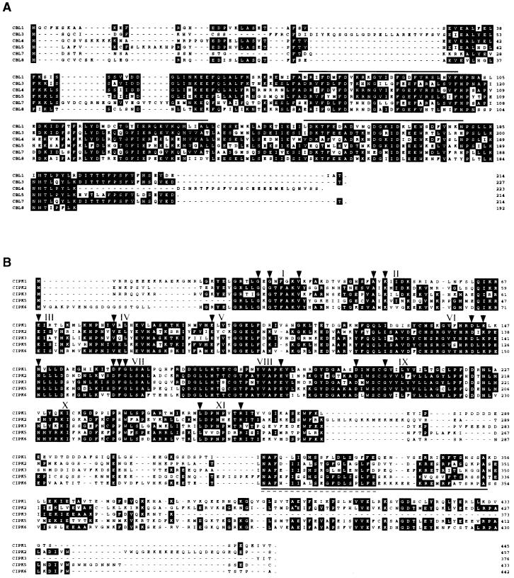
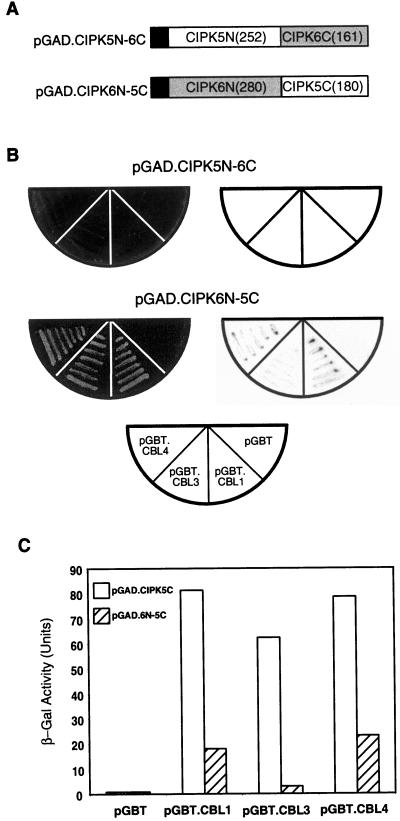
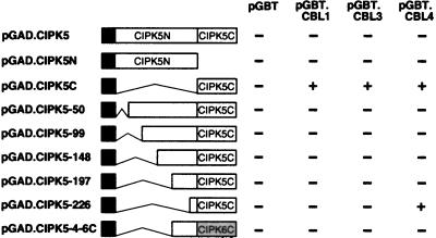
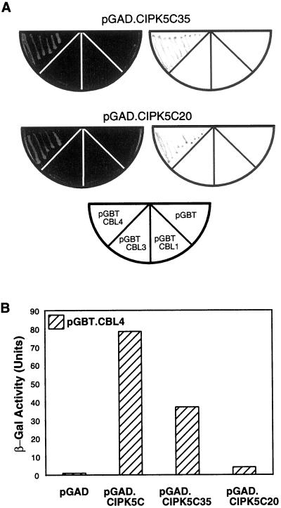
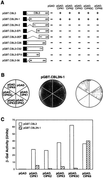
Calcium is a critical component in a number of plant signal transduction pathways. A new family of calcium sensors called calcineurin B-like proteins (AtCBLs) have been recently identified from Arabidopsis. These calcium sensors have been shown to interact with a family of protein kinases (CIPKs). Here we report that each individual member of AtCBL family specifically interacts with a subset of CIPKs and present structural basis for the interaction and for the specificity underlying these interactions. Although the C-terminal region of CIPKs is responsible for interaction with AtCBLs, the N-terminal region of CIPKs is also involved in determining the specificity of such interaction. We have also shown that all three EF-hand motifs in AtCBL members are required for the interaction with CIPKs. Several AtCBL members failed to interact with any of the CIPKs presented in this study, suggesting that these AtCBL members either have other CIPKs as targets or they target distinct proteins other than CIPKs. These results may provide structural basis for the functional specificity of CBL family of calcium sensors and their targets.


Figures

Figure 1
Figure 1
Sequence analysis of AtCBLs and CIPKs. A, Amino acid sequence comparison of AtCBL1 (GenBank accession no. AF076251), AtCBL3 (AF076253), AtCBL4 (Y18870), AtCBL5 (AC009519.4), AtCBL7 (AL078465.1), and AtCBL8 (AF069300.1). Solid lines above the sequence indicate the EF-hand motifs. Residues with black background indicate amino acids conserved in at least three genes, and dashes represent gaps to maximize alignment. B, Amino acid sequence comparison of CIPK1 (GenBank accession no. AB022219), CIPK2 (AF286050), CIPK3 (AF286051), CIPK5 (AF285105), and CIPK6 (AF285106). Arrowheads and Roman numerals above the sequences indicate the conserved amino acids and subdomains of Ser/Thr protein kinases, respectively. Solid and dashed underlines indicate regions used to create pGAD.CIPK5C35 and PGAD.CIPK5C20 plasmids, respectively.
Figure 2
Figure 2
The kinase domain of CIPK5 interferes with the interaction between AtCBLs and the C-terminal region of CIPKs. A, Schematic diagram of the chimeric constructs. The pGAD.CIPK5N-6C and pGAD.CIPK6N-5C plasmids were created as described in “Materials and Methods.” White and light gray boxes represent CIPK5 and CIPK6, respectively. B, Yeast two-hybrid assays. The chimeric plasmids were transformed into the Y190 yeast cells, which contain each of the pGAD plasmids indicated in the half circle at the bottom. The half circles at left indicate growth of the yeast cells on SC-His-Leu-Trp medium. The half circles at right show the filter-lift assay. C, Measurement of β-galactosidase activity. Three individual transformants were used to measure the β-galactosidase activity as described in “Materials and Methods.” Each value represents the average.
Figure 3
Figure 3
Identification of inhibitory domain in the N-terminal region of CIPK5. A series of N-terminal deletion mutants of CIPK5 were cloned into the pGAD vector and transformed into Y190 yeast cells containing pGBT, pGBT.CBL1, pGBT.CBL3, or pGBT.CBL4. Co-transformed yeasts were assayed for bait-prey interactions by determining their growth and measuring β-galactosidase activity. The plus signs indicate both yeast growth and color development in the filter-lift assay. The minus signs represent no growth. Other symbols are the same as in Figure 2.
Figure 4
Figure 4
The small conserved domain in the C-terminal region of CIPK5 interacts with AtCBL4 at reduced strength. A, Yeast growth and filter-lift assays. The pGAD.CIPK5C35 and pGAD.CIPK5C20 plasmids were created as described in “Materials and Methods.” The plasmids were then co-transformed into the Y190 yeast strains with pGBT, pGBT.CBL1, pGBT.CBL3, and pGBT.CBL4. The half circles at left indicate yeast growth on the selection medium. The half circles at right show the filter-lift assay results. The half circle at bottom represents the arrangement of the yeast strains containing the indicated plasmids. B, Measurement of β-galactosidase activity from the yeast strains containing pGBT.CBL4 and the indicated pGAD plasmids. Three individual transformants were used to measure the β-galactosidase activity as described in “Materials and Methods.” Each value represents the average.
Figure 5
Figure 5
Small deletions in either N- or C-terminal ends of AtCBL3 dramatically changes its ability to interact with CIPKs. A, Yeast growth on the selection medium. Solid black boxes indicate the DNA-binding domain in the pGBT vector. Numbers in the open boxes represent the beginning and the ending positions of AtCBL3 protein fragments. Each deletion construct was transformed into the Y190 yeast strains containing pGAD, pGAD.CIPK1, pGAD.CIPK2, pGAD.CIPK3, pGAD.CIPK5C, and pGAD.CIPK6, respectively. Yeast growth was monitored on the selection medium and indicated as growth (+) and no growth (−). B, Filter-lift assay of pGBT.CBL3N-1. The pGBT.CBL3N-1 plasmid was transformed into the yeast cells, which contain each of the indicated pGAD plasmids (left circle). The circles in the middle and at right show the results of yeast growth and filter-lift assay, respectively. C, Measurement of β-galactosidase activity. Three individual transformants were used to measure the β-galactosidase activity as described in “Materials and Methods.” Each value represents the average.

References

    1. Ames JB, Ishima R, Tanaka T, Gordon JI, Stryer L, Ikura M. Molecular mechanics of calcium-myristoyl switches. Nature. 1997;389:198–202. - PubMed
    1. Braunewell K-H, Gundelfinger ED. Intracellular neuronal calcium sensor proteins: a family of EF-hand calcium binding proteins in search of a function. Cell Tissue Res. 1999;295:1–12. - PubMed
    1. Breeden L, Nasmyth K. Regulation of the yeast HO gene. Cold Spring Harbor Symp Quant Biol. 1985;50:643–650. - PubMed
    1. Bush DS. Calcium regulation in plant cells and its role in signaling. Annu Rev Plant Physiol Plant Mol Biol. 1995;46:95–122.
    1. Calvert PD, Klenchin VA, Bownds MD. Rhodopsin kinase inhibition by recoverin: function of recoverin myristoylation. J Biol Chem. 1995;270:24127–24129. - PubMed

MeSH terms

Associated data