. 2000 Dec;14(12):1962-75.
doi: 10.1210/mend.14.12.0575.

Activation of peroxisome proliferator-activated receptors (PPARs) by their ligands and protein kinase A activators

Affiliations

Activation of peroxisome proliferator-activated receptors (PPARs) by their ligands and protein kinase A activators

G Lazennec et al. Mol Endocrinol. 2000 Dec.

Abstract

The nuclear peroxisome proliferator-activated receptors (PPARs) alpha, beta, and gamma activate the transcription of multiple genes involved in lipid metabolism. Several natural and synthetic ligands have been identified for each PPAR isotype but little is known about the phosphorylation state of these receptors. We show here that activators of protein kinase A (PKA) can enhance mouse PPAR activity in the absence and the presence of exogenous ligands in transient transfection experiments. Activation function 1 (AF-1) of PPARs was dispensable for transcriptional enhancement, whereas activation function 2 (AF-2) was required for this effect. We also show that several domains of PPAR can be phosphorylated by PKA in vitro. Moreover, gel retardation experiments suggest that PKA stabilizes binding of the liganded PPAR to DNA. PKA inhibitors decreased not only the kinase-dependent induction of PPARs but also their ligand-dependent induction, suggesting an interaction between both pathways that leads to maximal transcriptional induction by PPARs. Moreover, comparing PPAR alpha knockout (KO) with PPAR alpha WT mice, we show that the expression of the acyl CoA oxidase (ACO) gene can be regulated by PKA-activated PPAR alpha in liver. These data demonstrate that the PKA pathway is an important modulator of PPAR activity, and we propose a model associating this pathway in the control of fatty acid beta-oxidation under conditions of fasting, stress, and exercise.


Figures

Fig 1
Fig 1. CT increases mPPARα activity on a PPRE containing promoter
Mouse PPARα was transfected along with TK-CAT or 2CYPA6-TK-CAT constructs in HEK-293 cells. Transfections were with 200 ng of TK-CAT or 2CYPA6-TK-CAT reporter construct and without or with 100 ng of pSG5-mPPARα expression vector per well. Cells were grown for 36 h in the presence of 1 μM of WY 14,643 (WY), with or without 1μg/ml CT. Results are shown as the mean ± SD (n = 6) of CAT activity after normalization for β-galactosidase activity.
Fig 2
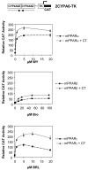
Fig 2. CT increases mPPAR activity at various concentrations of PPAR ligands
Mouse PPARα, β and γ were tested for their ability to respond to CT in HEK-293 cells. Transfections were with 200 ng of 2CYPA6-TK-CAT reporter construct and 100 ng of pSG5-mPPARα, β and γ expression vectors per well. Cells were grown for 36 h in the presence of various concentrations of WY 14,643 (WY), Bromopalmitate (Bro) and BRL 49,653 (BRL), with or without 1μg/ml CT. Results are shown as the mean ± SD (n = 6) of CAT activity after normalization for β-galactosidase activity.
Fig 3
Fig 3. PKA enhancement of PPAR activity via the ACO PPRE
Mouse PPARα, β and γ cDNAs were cotransfected in HEK-293 cells with ACO-TK-CAT reporter construct instead of the 2CYPA6-TK-CAT reporter construct in the same conditions as in fig 1. Cells were grown for 36 h in the presence of 1 μM WY 14,643 (WY), 50 μM Bromopalmitate (Bro) and 5 μM BRL 49,653 (BRL), with 1μg/ml CT when indicated. Results are shown as the mean ± SD (n = 6) of CAT activity after normalization for β-galactosidase activity.
Fig 4
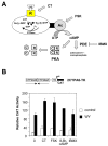
Fig 4. various PKA activators stimulate PPAR activity
A. Schematic representation of the PKA pathway. cAMP is produced from ATP by a membrane-bound adenylate cyclase (Ac). Transmembrane receptors (R) for numerous hormones, neurotransmitters and other stimuli (H) are coupled to adenylate cyclase via heterotrimeric G-proteins (α, β and γ subunits). This interaction promotes the exchange of GDP, bound to the α-subunit, for GTP and the subsequent dissociation of the α subunit form the βγ heterodimer. The Gα-GTP then binds to adenylate cyclase and modulates its activity. PDE: phosphodiesterase; FSK: forskolin; CT: cholera toxin;. B. 100 ng of pSG5-mPPARα was cotransfected in HEK-293 cells with 2CYPA6-TK-CAT reporter construct under the same conditions as in Fig 1. Cells were grown for 36 h in the presence of 1 μM of WY 14,643 (WY), with 1μg/ml Cholera toxin (CT), 50 mM 8-BrcAMP, 200 μM forskolin (FSK) or 10 μM IBMX when indicated. Results are shown as the mean ± SD (n = 3) of CAT activity after normalization for β-galactosidase activity.
Fig 5
Fig 5. PKA inhibitors can repress PPAR activity
A. Mouse PPARα, β and γ were tested for their ability to respond to CT in HEK-293 cells using 200 ng of 2CYPA6-TK-CAT reporter construct and 100 ng of pSG5-PPAR expression vectors. After lipofection, 10 μM of H89 was added to the medium 1 h before ligands. Cells were grown for 36 h in the presence of 1 μM WY 14,643 (WY), 50 μM Bromopalmitate (Bro) and 5 μM BRL 49,653 (BRL), with 1μg/ml CT when indicated. Results are shown as the mean ± SD (n = 6) of CAT activity after normalization for β-galactosidase activity. B. 30 μg of whole cell extracts from transfected cells were loaded on SDS-PAGE and probed by western blot using mPPARα antibody. The first lane corresponds to HEK-293 cells transfected with the empty pSG5 vector and the remaining lanes correspond to 293 cells transfected with pSG5-mPPARα vector and treated or not (−) with WY, CT or H89 under the same conditions as in Fig. 5A. C. 5 μg of the same WCE were used in gel shift assays using the ACoA probe.
Fig 6
Fig 6. RXR modulates PPAR activity in the presence of PKA activators
A. 100 ng of pSG5, pSG5-mPPARα and pSG5-mRXRβ2 expression vectors per well in combination or alone were cotransfected in HEK-293 cells with 200 ng of 2CYPA6-TK-CAT reporter construct. After lipofection, cells were grown for 36 h with or without 1 μM of WY 14,643 (WY), and 1 μM 9-cis retinoic acid (RA), with 1μg/ml CT when indicated. Results are shown as the mean ± SD (n = 6) of CAT activity after normalization for β-galactosidase activity. B. 100 ng of pSG5 or pSG5-mRXRβ2 expression vectors were cotransfected in HEK-293 cells with 200 ng of DR1-TK-CAT reporter construct per well. After lipofection, cells were grown for 36 h with or without 1 μM 9-cis retinoic acid (RA) and 1μg/ml CT when indicated. Results are shown as the mean ± SD (n = 6) of CAT activity after normalization for β-galactosidase activity.
Fig 7
Fig 7. The AB domain is dispensable for PPAR response to PKA
A. 100 ng of pSG5-mPPARα WT, or pSG5-mPPARα ΔAB, pSG5-mPPARα ΔLBD, or pSG5-mPPARα ΔAF2 constructs were transfected in HEK-293 cells with 200 ng of 2CYPA6-TK-CAT reporter construct per well. After lipofection, cells were grown for 36 h with or without 1 μM of WY 14,643 (WY) with or without 1μg/ml CT. Results are shown as the mean ± SD (n = 6) of CAT activity after normalization for β-galactosidase activity. B. Equivalent amounts of in vitro translated mPPARα WT, mPPARα ΔAB, mPPARα ΔLBD, or mPPARα ΔAF2 receptors were used in gel shift assays in combination with the ACoA probe. Lane 1 corresponds the probe alone and lane C to mock lysate. In addition, mouse RXRβ2 Sf9 cellular extract was eventually added (RXR). The arrow indicated the position of RXR retarded complex and the star the position of a non specific complexes. C. 100 ng of GAL, GAL-AF1, or GAL-AF2 constructs were transfected in HEK-293 cells with 200 ng of pG5CAT reporter construct. After lipofection, cells were grown for 36 h with or without 1 μM of WY 14,643 (WY) with or without 1μg/ml CT. Results are shown as the mean ± SD (n = 6) of CAT activity after normalization for β-galactosidase activity.
Fig 8
Fig 8. PPAR phosphorylation in response to PKA occurs mainly in the DBD
A. Regions encoding AB (ABα), DBD (DBDα) and LBD (LBDα) domains of mPPARα as well as DBD (DBDβ) and LBD (LBDβ) of mPPARβ were cloned in pGEX1 prokaryotic expression vector as GST fusions. In vitro PKA assay was performed as described in the Materials and Methods. The upper panel corresponds to the gel autoradiogramm and the lower panel to the protein expression levels after coomassie blue staining of the gel. The arrow labelled NS corresponds to a non specific band. B. Mapping of the main putative PKA sites of phosphorylation on mPPARα.
Fig 9
Fig 9. PKA modulates some PPAR target genes
A. Hepatocytes were isolated from wild-type and PPARα KO mice and cultured in Williams medium supplemented with 10% CDFCS. Cells were then treated for 24 h with or without 10 μM WY 14,643 (WY) and 1 μg/ml cholera toxin (CT). When H89 was used (H: 10 μM), it was added to the medium 1 h before treatment with WY or CT. After RNA extraction, 10 μg of total RNA were used for northern blotting and then hybridized sequentially with ACO, FABP and 28S probes. The figure shows a representative experiment. B. General model of cross-talk between fatty acids and PKA signalling involving PPARs. TG: triglycerides, FA: fatty acids, Glucocor: glucocorticoids, β OX: β oxidation, LPL: lipoprotein lipase.

Similar articles

Cited by

References

    1. A unified nomenclature system for the nuclear receptor superfamily. Cell. 1999;97:161–3. [letter] - PubMed
    1. Desvergne B, Wahli W. Peroxisome proliferator-activated receptors: nuclear control of metabolism. Endocr Rev. 1999;20:649–88. - PubMed
    1. Beato M, Herrlich P, Schutz G. Steroid hormone receptors: many actors in search of a plot. Cell. 1995;83:851–7. - PubMed
    1. Mangelsdorf DJ, Thummel C, Beato M, Herrlich P, Schutz G, Umesono K, Blumberg B, Kastner P, Mark M, Chambon P, et al. The nuclear receptor superfamily: the second decade. Cell. 1995;83:835–9. - PMC - PubMed
    1. Issemann I, Green S. Activation of a member of the steroid hormone receptor superfamily by peroxisome proliferators. Nature. 1990;347:645–50. - PubMed

Publication types

MeSH terms