. 2000 Dec 15;290(5499):2155-9.
doi: 10.1126/science.290.5499.2155.

Functional requirement for class I MHC in CNS development and plasticity

Affiliations

Functional requirement for class I MHC in CNS development and plasticity

G S Huh et al. Science. .

Abstract

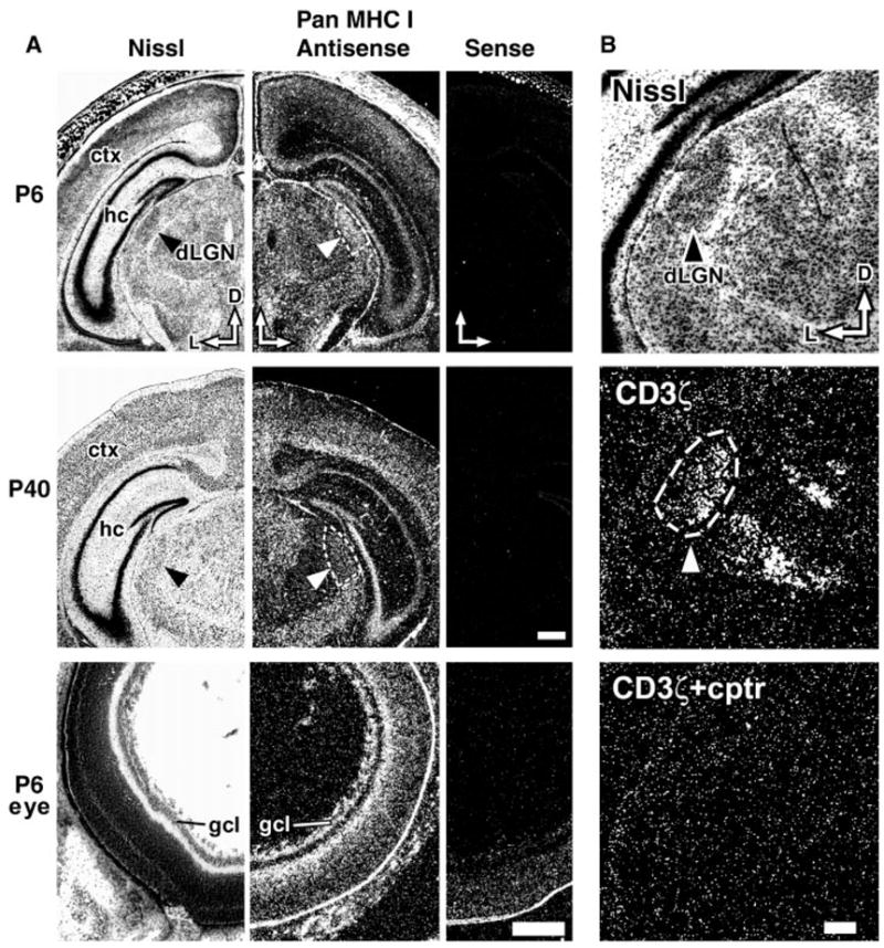
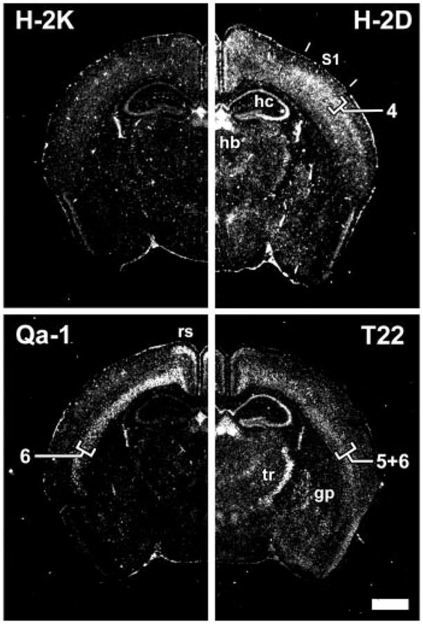
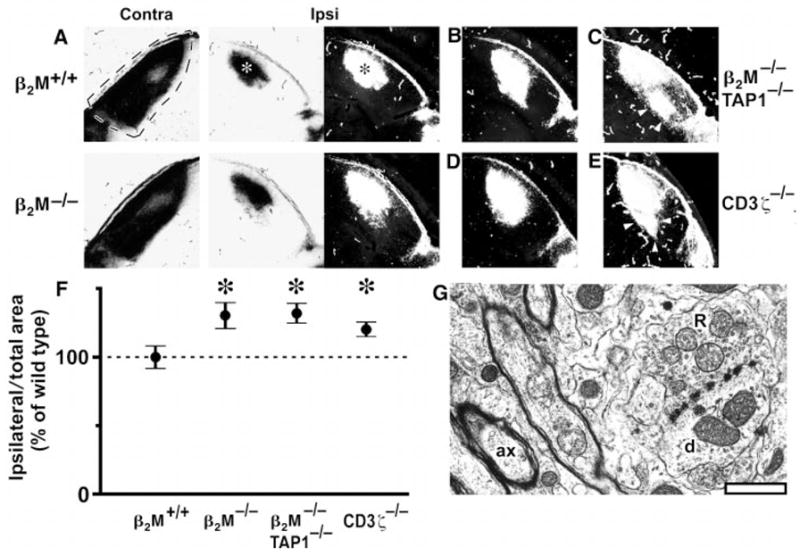
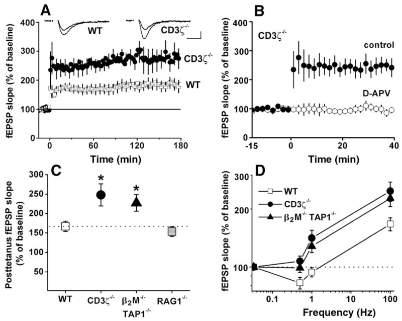
Class I major histocompatibility complex (class I MHC) molecules, known to be important for immune responses to antigen, are expressed also by neurons that undergo activity-dependent, long-term structural and synaptic modifications. Here, we show that in mice genetically deficient for cell surface class I MHC or for a class I MHC receptor component, CD3zeta, refinement of connections between retina and central targets during development is incomplete. In the hippocampus of adult mutants, N-methyl-D-aspartate receptor-dependent long-term potentiation (LTP) is enhanced, and long-term depression (LTD) is absent. Specific class I MHC messenger RNAs are expressed by distinct mosaics of neurons, reflecting a potential for diverse neuronal functions. These results demonstrate an important role for these molecules in the activity-dependent remodeling and plasticity of connections in the developing and mature mammalian central nervous system (CNS).


Figures

Fig. 1
Fig. 1
Class I MHC expression in mouse CNS. (A) Expression of class I MHC transcripts in coronal sections of the mouse CNS at P6 and P40 and in a cross section of P6 eye (13). Left column, adjacent Nissl-stained section; middle column, hybridization with antisense riboprobe under dark-field optics; right column, hybridization with control sense probe. D, dorsal; L, lateral; hc, hippocampus; ctx, neocortex; gcl, ganglion cell layer. Arrowheads and dashed lines indicate dLGN. Scale bar for P6 and P40 brains, 0.5 mm; scale bar for P6 eye, 250 μm. (B) Expression of CD3ζ in the dLGN during eye-specific layer formation. Upper panel, adjacent Nissl-stained coronal section of P6 mouse brain (arrowhead, dLGN). Middle panel, hybridization with CD3ζ antisense probe (dashed lines, dLGN); hybridization is also present in the ventroposterior nucleus of thalamus (down and to right of dLGN). Lower panel (cptr), excess of unlabeled competitor probe. Scale bar, 200 μm.
Fig. 2
Fig. 2
Expression of multiple class I MHC subclasses in distinct regions of the mature CNS. Coronal sections of P40 mouse brain analyzed by in situ hybridization, using subclass-specific probes indicated at top of each panel (13). S1, somatosensory cortex; hb, habenula; hc, hippocampus; rs, retrosplenial cortex; tr, thalamic reticular nucleus; gp, globus pallidus. Numerals (4, 6, 5+6) indicate neocortical layers. Scale bar, 1 mm.
Fig. 3
Fig. 3
Abnormal retinogeniculate projections but normal dLGN ultrastructure in mice deficient in class I MHC signaling. At P12, one eye was injected with WGA-HRP (23); after 1 day, anterograde axonal transport results in labeling of the entire retinal projection to the LGN. Labeling pattern in the dLGN is shown in bright-field optics (label is black) or as dark-field composites [label is white; see (24)]. (A) Representative projection from retina to dLGN contralateral (dashed lines; coronal section; dorsal is up; lateral is left) or ipsilateral to eye injected with WGA-HRP (asterisks indicate labeled area from ipsilateral eye: lateral is to right) in a P13 β2M+/+ wild-type mouse and a β2M−/− mutant mouse. (B and C) Representative (B) and extreme (C) examples of the projection from the ipsilateral eye observed in β2M−/−TAP1−/− mice. (D and E) Representative (D) and extreme (E) examples of the projection in CD3ζ −/− mice. Arrowheads indicate ectopic projections, which appear extensive under the more sensitive dark-field optics. Scale bar, 200 μm. (F) Graph of areas (±SEM) occupied by the ipsilateral retinal projection to the LGN for β2M+/+ (wild-type), β2M−/−, β2M −/−TAP1−/−, and CD3ζ−/− mice (24), normalized to total dLGN area. The ipsilateral projection area in β2M+/+ animals is set as 100% (horizontal dashed line). Asterisks indicate significant differences from β2M+/+ mice (P < 0.05, Student’s two-tailed t test). (G), Electron micrograph of the dLGN from a β2M−/−TAP1−/− mouse (at P24), showing a typical R-type synaptic bouton (R) making contacts with a dendrite (d). A well-myelinated axon (ax) is also present in this field. Scale bar, 1 μm.
Fig. 4
Fig. 4
Enhanced hippocampal LTP in mice deficient either for cell surface class I MHC expression or for CD3ζ. (A) Field EPSP (fEPSP) slopes in wild-type versus CD3ζ −/−-deficient mice. Tetanus was applied at time 0. (Insets) Superimposed sample fEPSPs recorded 10 min before or 180 min after tetanic stimulation from individual wild-type (left) and CD3ζ−/− (right) slices. Scale bar, 10 msec/0.25 mV. (B) NMDA receptor dependence of LTP in CD3ζ-deficient mice. Tetanus was applied at time 0 either in the absence [filled circles; from (A)] or presence (hollow circles) of 50 μM D-APV. All points in (A) and (B) are averages of four consecutive fEPSPs (means ± SEM, normalized to 15-min baseline) recorded from CA1. (C) Graphs summarizing degree of potentiation in wild-type, β2M−/−TAP1−/−, CD3ζ−/−, or RAG1−/− mice after 100-Hz tetanus. Data are shown for mice with histologically normal brains (48). Asterisks indicate significant differences from wild type (one-way ANOVA, P < 0.05). (D) Relation (logarithmic plot) between synaptic enhancement and stimulation frequency. Points at 0.033 Hz (test pulse frequency) indicate baseline values (horizontal dashed line). Points at 100 Hz are taken from (C). Values in (C) and (D) are mean fEPSP slopes for each genotype over the 1-hour period following tetanus. See text and (34) for methods.

Comment in

References

    1. Goodman CS, Shatz CJ. Cell. 1993;72:77. - PubMed
    1. Katz LC, Shatz CJ. Science. 1996;274:1133. - PubMed
    1. Crair MC. Curr Opin Neurobiol. 1999;9:88. - PubMed
    1. Wong RO. Annu Rev Neurosci. 1999;22:29. - PubMed
    1. Shatz CJ. Neuron. 1990;5:745. - PubMed

Publication types

MeSH terms