. 2000 Dec 15;19(24):6704-12.
doi: 10.1093/emboj/19.24.6704.

In vivo kinetics of protein targeting to the endoplasmic reticulum determined by site-specific phosphorylation

Affiliations

In vivo kinetics of protein targeting to the endoplasmic reticulum determined by site-specific phosphorylation

V Goder et al. EMBO J. .

Abstract

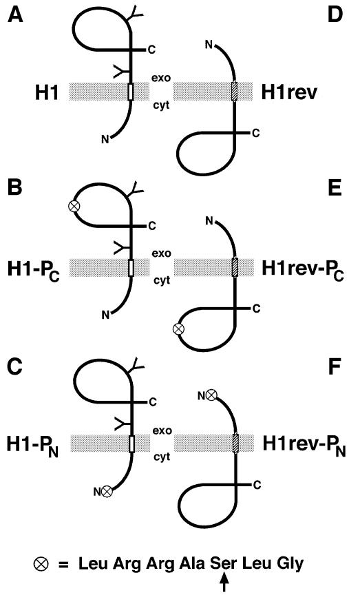
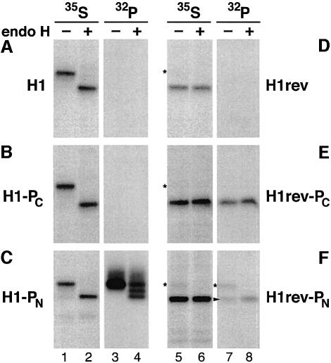
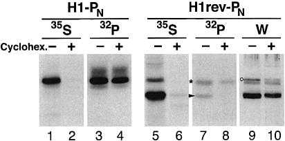
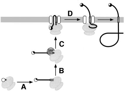
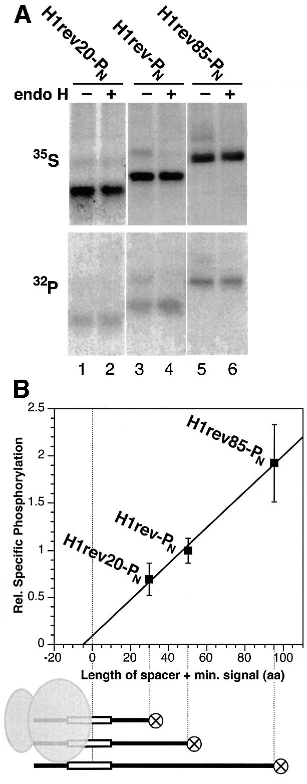
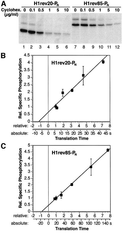
We have developed a novel assay to detect the cytosolic localization of protein domains by inserting a short consensus sequence for phosphorylation by protein kinase A. In transfected COS-1 cells, this sequence was labeled efficiently with [(32)P]phosphate only when exposed to the cytosol and not when translocated into the lumen of the endoplasmic reticulum. The phosphorylation state of this sequence can therefore be used to determine the topology of membrane proteins. This assay is sufficiently sensitive to detect even the transient cytosolic exposure of the N-terminal domain of a membrane protein with a reverse signal-anchor sequence. The extent of phosphorylation per newly synthesized polypeptide was shown to reflect the time of exposure to the cytosol, which depends on translation, targeting and translocation of the N-terminus. By altering the length of the N-terminal domain or manipulating the translation rate, it was determined that protein targeting is rapid and requires only a few seconds. The rate of N-terminal translocation was estimated to be approximately 1.6 times the rate of translation.


Figures

None
Fig. 1. Schematic representation of the model membrane proteins analyzed. H1 (A), H1-PC (B) and H1-PN (C) are Ncyt/Cexo proteins with a type II signal-anchor sequence (open rectangle) and a glycosylated (Y) C-terminal domain. H1rev (D), H1rev-PC (E) and H1rev-PN (F) contain a reverse signal-anchor sequence (hatched rectangle) and insert to ∼95% as unglycosylated Nexo/Ccyt proteins. The circle symbolizes the consensus sequence for phosphorylation by PKA shown below (with the target serine indicated by an arrow).
None
Fig. 2. Expression and phosphorylation pattern of the model proteins (AF) with opposite orientations. Transfected COS-1 cells were labeled with [35S]methionine or [32P]phosphate for 40 min and subjected to immunoprecipitation. Samples were incubated with (+) or without (–) endo H and analyzed by SDS–gel electrophoresis and fluorography. The asterisks indicate the position of a small percentage of proteins with a reverse signal-anchor that nevertheless inserted as type II (Ncyt/Cexo) membrane proteins. The arrowhead points to the labeled Nexo/Ccyt proteins. All 35S-samples and 32P-samples were exposed identically.
None
Fig. 3. Co-translational phosphorylation of H1rev-PN. COS-1 cells transfected with H1-PN or H1rev-PN were incubated with (+) or without (–) 100 µg/ml cycloheximide for 2 h before and 40 min during labeling with [35S]methionine or [32P]phosphate. After saponin extraction, the membrane proteins were immunoprecipitated and analyzed by SDS–gel electrophoresis and fluorography. The positions of phosphorylated H1rev-PN with Ncyt/Cexo and Nexo/Ccyt orientation are indicated by an asterisk and an arrowhead, respectively. To visualize the total H1rev-PN in the transfected cells, western analysis was performed on a parallel sample (W). A non-specific band detected upon western analysis is indicated by a circle.
None
Fig. 4. The phosphorylation tag acts as a timer of cytosolic exposure. The time of cytosolic exposure of the N-terminal phosphorylation sequence is determined by the time of translation of the N-terminal domain from the appearance of the tag until the signal has emerged sufficiently for interaction with SRP (A), the kinetics of SRP binding (B) and of targeting of the complex to the ER (C), and by the time required to translocate the N-terminal domain across the membrane (D). The phosphorylation tag is shown as a circle and its average modification is indicated by its filled portion.
None
Fig. 5. Specific phosphorylation as a function of the spacer length between the phosphorylation tag and the signal sequence. (A) H1rev20-PN, H1rev-PN and H1rev85-PN, which have spacers of 20, 40 and 85 residues, respectively, were expressed in COS-1 cells, labeled with [35S]methionine or [32P]phosphate, extracted with saponin, immunoprecipitated and, after incubation with (+) or without (–) endo H, analyzed by gel electrophoresis and autoradiography. Incorporation of 32P in the unglycosylated Nexo/Ccyt form of the proteins increased relative to the incorporation of 35S with increasing spacer length. (B) The specific phosphorylation ([32P]/[35S]) was quantified for three series of co-labeling experiments with a total of 10 samples for each construct, normalized to the specific phosphorylation of H1rev-PN, and plotted against the length of the spacer plus the minimal size of the signal to be recognized by SRP (∼10 residues, as illustrated below the graph).
None
Fig. 6. Specific phosphorylation as a function of translation rate. (A) Elongation was reduced by incubation of transfected COS-1 cells with different concentrations of cycloheximide during 40 min of [35S]methionine labeling. Saponin extraction, immunoprecipitation and fluorography were then performed. (B and C) COS-1 cells expressing H1rev20-PN or H1rev85-PN were labeled with [35S]methionine and [32P]phosphate, saponin extracted, immunoprecipitated and analyzed by gel electrophoresis. The specific phosphorylation (average with standard deviation from three determinations each) normalized to that determined in the absence of inhibitor is plotted against the relative translation time of the N-terminal domain from the emergence of the phosphorylation tag to the appearance of a minimal signal (translation of 30 and 95 residues, respectively). The lower scale provides absolute times of translation for a translation rate of 5 amino acids/s.

References

    1. Beltzer J.P., Fiedler,K., Fuhrer,C., Geffen,I., Handschin,C., Wessels,H.P. and Spiess,M. (1991) Charged residues are major determinants of the transmembrane orientation of a signal-anchor sequence. J. Biol. Chem., 266, 973–978. - PubMed
    1. Dale H. and Krebs,M.P. (1999) Membrane insertion kinetics of a protein domain in vivo. The bacterioopsin N terminus inserts co-translationally. J. Biol. Chem., 274, 22693–22698. - PubMed
    1. Dale H., Angevine,C.M. and Krebs,M.P. (2000) Ordered membrane insertion of an archaeal opsin in vivo. Proc. Natl Acad. Sci. USA, 97, 7847–7852. - PMC - PubMed
    1. Denzer A.J., Nabholz,C.E. and Spiess,M. (1995) Transmembrane orientation of signal-anchor proteins is affected by the folding state but not the size of the N-terminal domain. EMBO J., 14, 6311–6317. - PMC - PubMed
    1. Ellis L., Clauser,E., Morgan,D.O., Edery,M., Roth,R.A. and Rutter,W.J. (1986) Replacement of insulin receptor tyrosine residues 1162 and 1163 compromises insulin-stimulated kinase activity and uptake of 2-deoxyglucose. Cell, 45, 721–732. - PubMed

Publication types

MeSH terms